Predation event between a Praying Mantis (Mantodea: sp.) and a sub-adult female of Anolis cusuco. Photo Credit – George Lonsdale
A natural history note published September 2019 in the journal SAURIA details an unusual observation of anolivory by a Praying Mantis. Specifically, it discusses an event involving the predation of a sub-adult female Anolis (Norops) cusuco.
Anolis cusuco owes its name to its type locality in the cloud-forest of Cusuco National Park, Honduras, and is a species endemic to the country. Few publications exist regarding the natural history of this species and much regarding its ecology, including its potential predators, remain unknown. While a small contribution, this observation describes the first, albeit somewhat unsuspecting predator for Anolis cusuco.
Victoria Pagano’s page from the crowd-funding platform Experiment
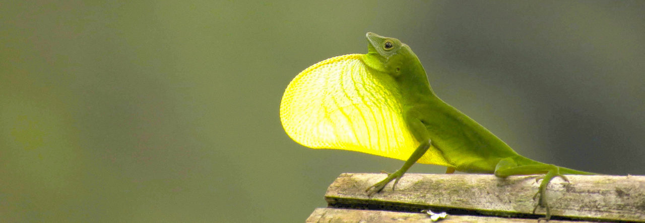
Green anoles (Anolis carolinensis) are talked about quite frequently here on Anole Annals, with 11 articles being published in 2018 and 2019 combined! As I am sure many of you are aware, green anoles change color from green to brown, and while it is known how, it is not yet known why. Although there have been multiple field studies into what causes green anoles to change color, the data have been inconclusive. This is why an experimental study is necessary to try to determine the cause of the color change.
In this experimental study, there will be two main hypotheses tested:
The first is the well known thermoregulation hypothesis. I will be testing this by establishing separate light and heat sources, and turning them on and off for different scenarios. If anoles change color for thermoregulation, then they would turn brown more frequently when the heat is off and the light is on.
The second hypothesis is the effect of increased stress. Stress will be induced by sliding a red disk towards the anoles multiple times at a high speed. Any color change that occurs within the red disk moving and the following 10 minutes will be documented as stress-induced.
I will not be able to test the advertisement signaling hypothesis due to feasibility. Because funding and space is limited, I do not have the capacity to house male anoles, as each one needs his own setup. Therefore, testing only females is the only feasible option, and by doing so, the advertisement signaling hypothesis will not be able to be tested, as this hypothesis pertains mainly to males.
To raise funding for this project, I am using an all or nothing crowdfunding platform called Experiment. As fellow anole lovers, I hope that you can help support my scientific endeavors by visiting my project page. All forms of support are greatly appreciated, from donations, to telling your friends about the project, or even by just reading my project page and commenting your thoughts! Whatever the contribution, I am very grateful, and am simply excited to be able to share what I am doing with all of you!
If you wish to learn more about this project, you can visit the project page, “What drives the color change in green anoles?”, where I have posted my methodology, protocols, and will be posting continuous updates on the progression of the project. If you become a contributor, you will have exclusive access to more updates, and will be able to learn more about the research.
My project page stops accepting donations on November 1st at 12:00 AM PT, so be sure to make your way over to the page by then to give your support!
Thank you for taking the time to read this article. I hope that you will explore the project page, and help support this cool and unique research!
An adult male Anolis amplisquamosus with black gorgetal scales immediately after capture (left); the same individual ~10 min later with white gorgetal scales. Photo Credit – John David Curlis
Anole dewlaps are excellent examples of a “complex signalling system.” They exhibit a staggering diversity of colours and patterns. Each dewlap is species specific and adapted to enable these lizards to communicate, attract mates and guard their territories from rivals or competitors. Generally, the colour of a dewlap (and its gorgetal scales) is considered an unchangeable descriptive trait. This colouration is not only relied upon by scientists looking to identify a species, but also by anoles that co-occur and partition with different species in their select niche.
Therefore, it might be surprising to learn that recent observations prove rapid colour change in anole gorgetal scales is possible. The question is, what implications does this have?
A recent publication in IRCF Reptiles & Amphibians details an observation of Anolis amplisquamosus whereby a male individual upon capture possessed black gorgetal scales that quickly changed to pale yellow. Upon consulting the literature, it seems only one prior documentation of colour change in gorgetal scales was reported (Leenders and Watkins-Colwell, 2003), coincidentally also involving a member of the same species clade.
This recent observation of chromatophoric regulation in anole gorgetal scales may be significant in the wider context of anole biology, in confirming photographically that coloration is not always a fixed descriptive or diagnostic feature — at least among members of the A. crassulus species group. Accordingly, this information suggests that some anoles may have the ability to regulate the colour of their gorgetal scales in the same manner as they regulate dorsal and lateral scale colour.
Because the colour of gorgetal scales is a character often used in species identification, understanding the mechanics and the purpose of such a change is crucial; as well as any implications to display behaviour, communication and anole interactions.
I recently received an email from Chris McMartin, the director of the Southwestern Center for Herpetological Research, about a population of brown anoles near his home in Montgomery County, Texas, just north of Houston. Chris has done a lot of preliminary research to understand how the Montgomery population is spreading, and would like to know how these lizards are related to the larger population in Harris County.
Interested? Keep reading!
With Chris’ permission, I’ve copied part of his email below:
“I’ve been casually (in my free time, mostly in the summer) researching Brown Anoles (Anolis sagrei) and their spread in southern Montgomery County where I live. As I amass observational data, I’ve noticed the lizards are abundant in some yards/neighborhoods, but nonexistent in adjacent yards/neighborhoods. I’m slowly trying to piece together additional factors (presence of outdoor cats, prevalence of certain landscaping features including decorative rocks and tropical plants, age of house/neighborhood, use of pesticides, etc.) which may explain not only the disparity in abundance but provide clues as to how to control their expansion.
One big question I have is whether the lizards are naturally expanding from a single introduction long ago (e.g. rapidly moving northward from Harris County, where they occur in densities many times higher than the highest I’ve observed in Montgomery County), or are an amalgamation of numerous discrete introductions (e.g. when a home installs new tropical plants from a nursery/home improvement store). Brown Anoles first showed up in my yard a little over a year ago, marking an expansion northward of about ¾ of a mile from my previous northernmost observations the year prior.
I have corresponded with Dr. Benson Morrill, who owns Rare Genetics Inc. offering DNA analysis for (at this time) colubrid snakes (primarily for sex determination) and inquired as to the possibility of sending him samples from various neighborhoods in my area in an effort to determine whether they represent a contiguous related population or are the result of discrete introductions. He says the process to conduct this analysis would be cost-prohibitive for a private individual such as myself, but perhaps a university student would like to take on the project.
As it is, I currently spend what surprisingly-little free time I have in the summer exploring neighborhoods in my neck of the woods and documenting my obervations—around 60 hours this past summer between field work and analysis—and I’m approaching my limit of resources in time (and definitely money, if considering DNA analysis as part of the project). This is where I think perhaps a graduate student might be interested in taking on a study of Brown Anoles as a thesis project…lots of possible threads to pull (competition with natives, rate of range expansion, effect of occasional hard freezes on population, etc.).
I’ve published articles in local magazines/newspapers about the lizards and have a public-opinion survey from a year ago (still awaiting analysis) trying to find any links between various conditions (age of neighborhood, presence of outdoor cats, etc.) and occurrence/prevalence of browns, especially with respect to A. carolinensis. Some interesting things seem to be occurring. Anecdotally, browns are eating greens (hatchlings), Broad-headed Skinks are eating browns, and greens are eating the skinks (with photographic evidence)! Sort of a three-way lacertilian arms race.”
If this sounds like just the opportunity you’ve been looking for, contact Chris at yall [at] mcmartinville.com.
This pale little lizard is one of the world’s first genetically edited non-avian reptiles. Image Credit: Courtesy of Ashley Rasys, University of Georgia
Compared to mammals, reptiles have a weird way of reproducing—and in the spring of 2017, that put Ashley Rasys in something of a pickle.
For months, the University of Georgia biologist was struggling to come up with a way to tinker with the genes of the brown anole (Anolis sagrei), a petite, pointy-faced lizard native to Cuba and the Bahamas.
The reptile had initially caught Rasys’ eye because of, well, its eyes. People with albinism often have poor vision due to problems with their foveae, the dense pits of cells at the back of the eyes that confer visual acuity. While foveae are lacking in most mammals, they’re present in lizards—making them intriguing candidates for studying the genes that impact foveal function.
There was just one problem: Reptiles aren’t easy to genetically manipulate. In other common laboratory animals, like mice and zebrafish, a tool called CRISPR has made DNA editing a breeze. The procedure typically involves injecting freshly fertilized eggs with gene-editing machinery, creating a change that would propagate when the cell divided.
But a few quirks ruled out that particular strategy in these lizards. Female anoles can store sperm for many months before fertilizing their eggs internally, making it difficult to time the introduction of the CRISPR cocktail. Anole fertilization also cues the formation of a soft, delicate eggshell that’s hard to penetrate without damaging the embryo.
That meant Rasys and her advisor, Doug Menke, had to get creative. So they decided to shift the injection back a developmental step, targeting eggs still maturing in the females’ ovaries. “At this point, they’re just hanging out in the lizard, waiting to be fertilized,” Rasys says.
Thanks to CRISPR gene editing, one of these brown anoles isn’t exactly brown. Image Credit: Courtesy of Ashley Rasys, University of Georgia
The procedure took more than a year to perfect. But in the fall of 2018, Rasys, Menke, and the rest of their team hatched the world’s first gene-edited non-avian reptile: a red-eyed albino anole with near-transparent skin. According to the team’s study, published today in the journal Cell Reports, its birth marks a breakthrough for the field of developmental genetics, and hints that similar experiments may be possible in some of the other 10,000-plus species of non-avian reptiles that scuttle the Earth.
“This technology is really important and exciting,” says Martha Muñoz, an evolutionary biologist and anole researcher at Yale University who was not involved in the study. “This really opens up the door for other groups to think outside of traditional model organisms [like mice and zebrafish]…the sky’s the limit.”
With albinism in mind, Rasys and her colleagues set out to mutate the anoles’ tyrosinase gene, which governs pigmentation and has been linked to foveal function in humans. Manipulating this gene, Rasys explains, also made for an easy marker of success: If the procedure ended up generating albino anoles down the line, they’d be pretty tough to miss.
After rounding up 21 female brown anoles from the wilds of Orlando, Florida, the researchers gently anesthetized the lizards and opened them up. In anoles, the ovaries are transparent, making it easy to eyeball their contents “like a train of developing eggs,” Menke says.
The team selected 146 of these growing eggs and injected them with the classic CRISPR recipe: a pair of molecular scissors and a series of DNA-binding “guides” that would show them where to cut—in this case, the tyrosinase gene.
The researchers then had to wait another three months or so for the females to fertilize and lay the eggs. And even when this generation hatched, they thought there’d likely be more work to do, Rasys says. Since the CRISPR concoction had been delivered to eggs that were later fertilized by unaltered sperm, the offspring were expected to be hybrids—half edited, half unedited. These lizards then would need to be bred further to yield albinos, which must inherit the mutation from both parents for the trait to manifest.
But as Rasys watched her first clutch of gene-edited eggs grow, she noticed something strange. About a week before they were due to hatch, most of the embryos had darkened from pink to gray—an indication that they’d started producing pigment. A handful, however, retained their initial pallor, even as they continued to swell in size.
A few days later, Rasys arrived at the lab to find a newly-hatched, inch-long albino, stretching its ghostly pink legs. “It was so exciting to see it,” she recalls. “I thought, ‘It’s so cute.’”
In total, four out of the team’s 146 CRISPR-injected embryos were obvious albinos, surprising the entire team. There’s no way to know exactly what happened, but Menke’s leading theory is that the CRISPR components remained active in some of the eggs long enough to work their magic on both the maternal and paternal copies of the tyrosinasegene.
In their native habitat, brown anoles can blend in pretty easily with tree bark. Such is not the case for albino mutants produced by CRISPR gene editing. Image Credit: Courtesy of Ashley Rasys, University of Georgia
Genetic screening revealed another five embryos to be the half-edited hybrids the team had initially expected. And when the researchers partnered one of these CRISPR mutts with an unmanipulated mate, the mutation was passed on to some of the pair’s offspring, suggesting the edited gene was heritable.
There’s still plenty of tinkering to do, Menke says. As they report in the study, the team’s gene-editing success rate was around 6 percent—a figure that pales in comparison to the near-perfect efficiency rates that have been reported in zebrafish and mice.
But just showing gene-editing is possible in this system is a big deal, says Ambika Kamath, a behavioral ecologist at the University of California, Berkeley who was not involved in the study. Albinism implications aside, anoles have long been studied by evolutionary biologists and ecologists. In their native Caribbean, the lizards have split into many lineages, but understanding this diversification “has primarily been a historical science…involving stitching together patterns that happened a long time ago,” Muñoz says. “By extending CRISPR to Anolis, we can now mechanistically test some [evolutionary] hypotheses.”
As more applications surface, however, “we don’t want to be releasing CRISPRed lizards into the wild willy-nilly,” Kamath says, without a better understanding of how these sorts of introductions would affect the population at large.
An albino anole produced by CRISPR gene editing (left) next to a typical brown anole. Image Credit: Courtesy of Ashley Rasys, University of Georgia
And it might be more than lizard lives at stake. Menke thinks the team’s technique is likely to work in a variety of reptiles, many of which share the anole’s mode of reproduction. There’s even the possibility, he says, that the method could be adapted for birds, which are cut from the same evolutionary cloth. Scientists have hatched CRISPant chicks in the past, but as in lizards, bird embryos are hard to pinpoint at the single-cell stage, making current editing procedures complex and laborious.
Carolyn Neuhaus, a bioethicist at the Hastings Center who was not involved in the study, cautions that as CRISPR continues to be debuted in more and more organisms, the how, when, and in whom of gene editing will need to remain transparent. Though many experiments—including the ones in this study—have the potential to advance science and human health, she says, technology like this shouldn’t be used in a new species “just because it’s there.”
“We rely on scientists to create accurate and reliable knowledge, and that’s a huge responsibility,” she says. “With the CRISPR craze…I just hope it happens as mindfully and carefully as possible.”
When I first encountered Anolis baleatus, this Hispaniolan crown-giant was mostly an inconvenience. At the time I was gathering data for my doctoral thesis by cycling preserved anoles through a µCT-scanner. Most of the adult specimens of A. baleatus were just too large to easily fit into the scan chamber, so it took a lot of patience and creativity to acquire any decent images of the appendicular girdles, which are the body parts I was interested in.
During that process I also acquired radiographic images of the head skeleton, and found unusual patterns of crenulation in this species. The cranium of Anolis baleatus displays a great degree of seemingly asymmetrical (or at least somewhat irregular) ornamentation across its dorsal surface. This is especially pronounced on the prefrontal and frontal bones, and completely obscures all superficial distinction between them in adult lizards. In adults, cranial ornamentation is also borne by the paired nasals, maxillae, and postorbitals, and the parietal (see figure).
Both Steven Poe (1998) and Susan
Evans (2008) mentioned this ossified garnish, but a thorough account of their
variation among anoles remains absent from the primary literature. Richard
Etheridge and Kevin de Queiroz (1988) were probably the first to report on skull
ornaments in anoles (as part of a discussion of several iguanian lizards with
similar cranial adornments), and remarked that the distribution patterns of
dermal rugae may reflect those of the topographically associated epidermal
scales.
Overall, this ornamentation appears
to be relatively uncommon among anoles, especially to the degree expressed in Anolis baleatus (and several other
crown-giant ecomorph anoles). Considering the osteologically robust appearance
of crown-giants, even at early stages of ontogenetic development, this gives
rise to questions regarding the development of these ornamental patterns.
Thanks to the collection efforts of Luke Mahler (University of Toronto), and a
postdoctoral position in his lab, I was able to acquire CT-image data
representing an ontogenetic series of this species, ranging from very young
juveniles to skeletally mature adults.
While parts of the paired frontals
of juveniles are covered in modest eminences, prominent cranial ornamentation
is absent from small specimens (see figure). Likely, growth of these ornaments
begins very late during ontogenetic development. Ornaments on the prefrontals
and parietal are only evident in specimens that, to the best of our judgement, are
approaching sexual maturity. We looked at fifteen specimens per sex,
representing a range of juvenile and subadult sizes, and this general pattern is
consistent throughout the image data. Schwartz (1974) inferred that anoles in
the ricordii group reach sexual
maturity between 100 and 110 mm snout-vent length (SVL), and we observed the
first prominent ornaments at sizes between 90 and 95 mm SVL. Assuming that
differences in size directly represent ontogenetic growth, these findings imply
that Anolisbaleatus starts to grow elaborate ornamentation as it approaches
sexual maturity, and that expansion and growth of these ornaments then
continues into skeletal maturity. Interestingly, both males and females appear
to develop them at roughly the same body size.
The function and evolutionary cause of these structures remain unknown, and these are questions we are currently investigating. Body size is an important correlate for the occurrence of cranial ornaments, but these structures may also conceivably play roles in defense, feeding, or intraspecific agonistic interactions. Stay tuned!
Videos
A. baleatus, female, 55 mm SVL
A. baleatus, female, 65 mm SVL
A. baleatus, female, 96 mm SVL
A. baleatus, female, 126 mm SVL
References
Etheridge, R. & de Queiroz, K. (1988): A phylogeny of Iguanidae.─ [In:] Estes, R.D. & Pregill, G.K. (eds.): Phylogenetic Relationships of the
Lizard Families: Essays Commemorating Charles L Camp, 283-367; Stanford:
Stanford University Press.
Evans, S. (2008): The skull of lizards and tuatara.─ [In:] Gans, C., Gaunt, A.S. &
Adler, K. (eds.), Biology of the Reptilia, vol. 20:1-347;
Society for the Study of Amphibians and Reptiles, Ithaca, New York.
Poe, S. (1998): Skull characters and the cladistic relationships of
the Hispaniolan dwarf twig Anolis.─ Herpetological Monographs, 12:192-236; The Herpetologists’ League.
Schwartz, A. (1974): An analysis of variation in the Hispaniolan
giant anole, Anolisricordi Dumeril and Bibron.─ Bull. Mus. Comp. Zool., 146:89-146.
Emma Higgins presenting her work at Island Biology 2019
The third meeting of the nascent Society for Island Biology took place recently in stunning La Reunion in the western Indian Ocean. Conference goers were treated to a wonderful venue at the Université de La Réunion in St. Denis, whose campus looks out down the gentle slope to the open sea. Four hundred attendees from around the world reinforced what we already knew— that island biology as a study attracts a large number of researchers from very diverse fields of study. The conference organizers also are leading the way on making our meetings in remote locations more responsible; using live streaming of the sessions meant that some interested scientists could skip the travel and stay home to watch the sessions. But the real asset was that the organizers calculated the air travel carbon footprint for all attendees, finding that ~30 hectares of forest would need to be planted to offset the carbon emissions. Happily, that is exactly what they did! The conference organizers and hosts, in partnership with communities and other organizations, committed to reforesting exactly that amount in La Réunion and Mauritius.
OK, now on to the anoles! Well, given the remoteness of the meeting location (nearly the antipode of the Caribbean*) perhaps it is not too surprising that few anologists attended. But I am happy to report that Emma Higgins did, and gave an excellent presentation on her work with anoles on the island of Utila, one of the Honduran Bay Islands.
Emma is a 3rd year PhD student in Adam Algar’s lab at the University of Nottingham, where her thesis is focused on using emerging technology to study lizard thermal biology under changing conditions (think development, climate change, and species introductions). When I say using emerging technology, I mean using #allthetech; Emma uses 3D printing, drones fitted with thermal cameras, Sentinel satellites, and LIDAR to generate her data! Her motivation follows from asking what factors control the abundance, distribution, and microhabitat of anoles on Utila, and whether these variables might be better estimated at extremely fine scales using emerging technology.
A bit of background, there are four species of anoles on Utila, including the endemics A. bicaorum and A. utilensis. An additional native species is A. sericeus, which also occurs elsewhere in the Bay Islands as well as on the mainland. The fourth species is everyone’s favorite— A. sagrei! Utila has experienced a surge of development in the last 10 years, with new roads and development going up faster than conservationists can keep track of. This is a major threat to the island wildlife, which includes an endemic iguana known as The Swamper (Ctenosaura bakeri) which favors the dwindling mangrove forests.
Emma’s work involves collecting data both at anole-level as well as above the canopy. She uses a DJI Phantom 4 drone platform fitted with a near-infrared camera to estimate a normalized difference vegetation index (NDVI, a measure of “greenness”) of the forest canopy across habitats, and found that just NDVI explains 28% of the spatial heterogeneity of lizard operative temperatures (in a mixed-model framework). This suggests that her drone can identify suitable thermal environments for lizards from above the canopy. I should mention that her resolution here is 4cm/pixel! She plans to zoom out to space and test whether similar imagery from the Sentinel 2 satellite will also be useful. Below the canopy, Emma is using LIDAR to simultaneously conduct forest shade modeling (for super fine-scale temporal variation in thermal microhabitat). LIDAR also detects perch availability, as it detects tree trunks very well. Emma also uses 3D printing to produce hundreds of anole models, each fitted with an iButton® temperature recorder and placed on perches in the forest. Each lizard print takes 52 minutes, so Emma ended up taking the printer to her flat to print 24/7 in preparation for her field season!
I should mention that Emma was joined at the conference by two other excellent scientists— lab mate Vanessa Cutts and fellow Utila lizard biologist Daisy Maryon, both of whom won awards for their posters at the conference!
Stay tuned for the announcement for the 2022 Island Biology meeting, to be held on either Mallorca in the Balearic Islands or Wellington, New Zealand. Also stay tuned for Emma’s results; we look forward to hearing more about her work!
Climate change is negatively affecting Squamates all over the world, and the perspectives for the next 50 years are worrisome. Although more than 200 studies were published in the last 20 years on this subject, only 23 present some information on Anoles, and none of them focused on population dynamics – until now. In the most recent issue of Comparative Biochemistry and Physiology Part A: Molecular & Integrative Physiology, Diele-Viegas et al. evaluated the possible effects of climate change on two populations of three species of lizards from the Brazilian Amazonia, including Norops fuscoauratus.
The idea of this research came from the first author, Dr. Luisa, during her PhD classes back in 2017. She was taking classes on population ecology and had the idea of to combine environmental thermal adequacy with the b-d model, which considers survival and reproductive rates to calculate population dynamics andevaluate the impact of climate change on population dynamics of her study objects, Amazonian lizards. She thus started to search in the literature for articles that focused on something like what she was thinking, and discovered that this had not been done before, at least not for squamate reptiles. She also noticed that data on Amazonian lizards’ life history is very scarce on literature, which could be a deterrent to her getting the job done. After speaking with her advisors (Dr. Fred Rocha and Dr. Fernanda Werneck), Luisa decided to estimate a tolerance index considering the relationship between the upper temperature limit of the animal activity restriction and the environmental temperature of the microhabitat in which this animal occurs and used this index as an approximation of the populations’ survival rates. This allowed her and her advisors (both coauthors of this article) to circumvent the data scarcity and put Luisa’s idea into practice, leading to the publication of this article.
Considering the tolerance index mentioned, the authors predicted that Norops fuscoauratus is likely to became locally extinct at one of the evaluated sites, Reserva Ducke, which is an almost urban reserve in the Amazonian heart. The hatchlings’ tolerance to environmental change was considered the most sensitive vital trait evaluated, highlighting the species’ vulnerability. Also, considering that the response to selection is likely to be too slow in anoles, an evolutionary change in N. fuscoauratus is unlikely to occur considering current rates of environmental change, which reinforces the species’ vulnerability at local scale. This study represents the first effort to evaluate population sensitivity to climate change among reptiles. The authors highlight the need for new studies focusing on this subject to provide theoretical and empirical basis for biologically informed conservation strategies and actions to avoid the extinction of several species around the world. Also, Luisa highlights that, as scientists, we should always value our ideas – our curiosity is the fuel that moves science into progress.