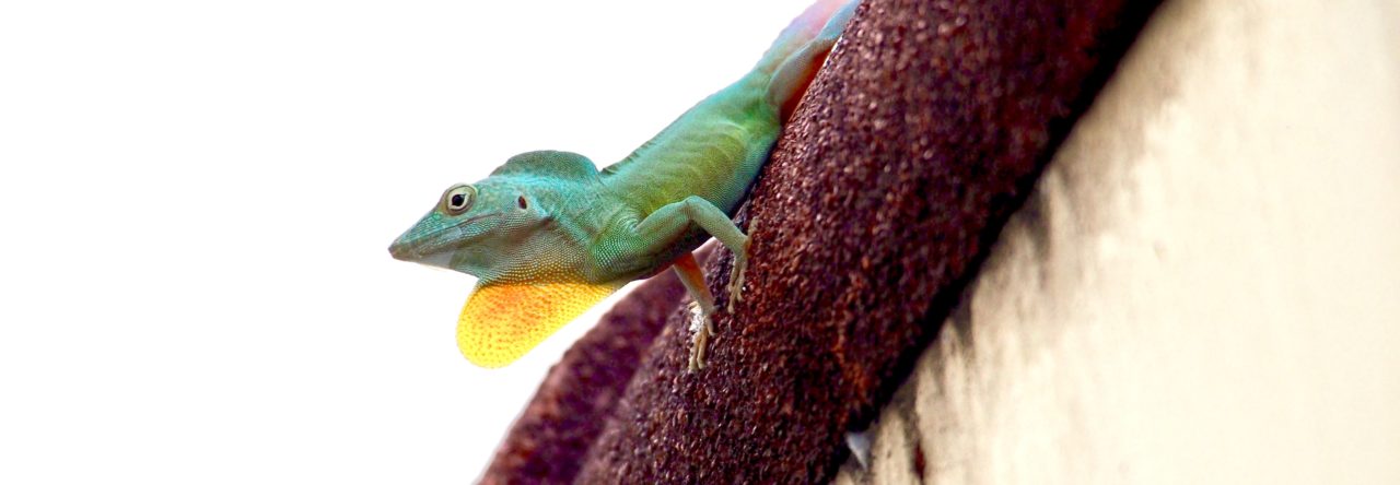
 I caught my first anole talk at this year’s Joint Meeting of Ichthyologists and Herpetologists in Chattanooga, Tennessee. James Stroud presented the results of work with Ken Feeley on modeling the niche of the brown anole (Anolis sagrei). Using data acquired from GBIF, Stroud showed that the environmental conditions experienced by brown anoles in their introduced range are outside of the environmental conditions experienced by brown anoles on Cuba. Stroud discussed how these data from the invasive range of the brown anole might be used to develop a more accurate model of this species’ fundamental niche. This is a work in progress.
There is considerable variation in phallus morphology among the major groups of amniotes (phallus used herein to be inclusive of both the penis and clitoris). Just for starters, while most clades – including mammals, birds, turtles, and crocodilians – have a single midline phallus, squamates have paired hemiphalluses. Although herpetologists have long appreciated morphological variation in the hemipenis for its systematic value, understanding the nuances of anatomical homology, homoplasy, and novelty at this larger scale has not been as widely addressed. Recently, the Cohn lab of the University of Florida (of which I am now a member) undertook this challenge from a developmental perspective, studying development of external genitalia in representatives of each reptilian clade: the ball python (Python regius), the pond slider (Trachemys scripta), three duck species, the American alligator (Alligator mississippiensis), and who else, but the green anole (Anolis carolinensis). A synthetic review of the complete series will have to wait for another post, but reprints of each paper are available on the lab’s website to hold over the most curious. But because of the growing interest in anole nether regions, I will briefly highlight the recent findings regarding hemiphallus development in the green anole.
The Wade lab has previously shown that both male and female green anoles develop similar hemiphalluses during the early stages of genital morphogenesis, which then later differentiate into sex-specific reproductive structures. Building upon this observation, Gredler et al. described the embryology of the green anole hemiphallus from the earliest stages of morphogenesis through sexual differentiation. Hemiphallus development begins around the time of oviposition when three sets of paired swellings appear between the cloaca and the developing hindlimb bud, reminiscent of what is observed in other amniote clades. These swellings expand and meet at the midline to form the external lips of the cloaca or remain lateral to the cloaca and mature into the hemiphalluses. Following morphogenesis, the male hemipenis continues to elongate as it forms its distinctive lobes and sulcus spermaticus while the female hemiclitores gradually regress into the cloaca. Further details of the developmental anatomy of internal reproductive structures and gene expression patterns of several key molecules associated with genital morphogenesis are described in the paper.

Fig. 4 of Gredler et al. illustrating sexual differentiation of the hemiphalluses. Red arrow highlights the formation of the sulcus spermaticus.
Although there is some variation among squamates in the relative timing of the emergence and fusion of the paired swellings associated with hemiphallus development, these results are largely consistent with classical embryological descriptions of squamate genitalia (summarized by Raynaud and Pieu in Biology of the Reptila volume 15). But the revival of this body of literature in a comparative and molecular context brings new research questions to our collective table. As discussed by Gredler et al., the seemingly modular relationship between the genital swellings, cloaca, and limb buds may be particularly interesting in the context of repeated body elongation and limb loss among squamates. Better understanding of the relationship between cloacal and phallus development may also shed new light on the mechanisms of reproductive isolation, the coevolution of male and female reproductive organs, and evolving patterns of sexual conflict. Furthermore, there remain open many mechanistic questions regarding the molecular patterning of the hemiphalluses and which processes are hormone dependent that can now be more thoroughly addressed using the newly available sex-specific molecular markers. Considering the growing literature on hemipenis variation and expanding access to genomic resources in Anolis, these may be particularly fruitful areas for future investigation.

Variation in the back patterns of Anolis sagrei in the Bahamas. From Calsbeek and Cox (2010).
The confusing conundrum of the polymorphic females continues. We’ve written about this phenomenon in previous posts [e.g., 1,2]. Within and between populations, female back patterns vary, including lines, stripes, diamonds, blotches, and nothing at all. What is the significance of this variation? In some cases, but not others, females with different patterns use different microhabitats–higher, wider, etc.
The latest contribution features work on the Bahamian island of Eleuthera, where three patterns co-occur. Writing in Herpetologica, Les et al. add a new twist–back pattern variants differ in hindlimb length. But they don’t differ in sprint speed (which is weakly correlated to body size and relative limb length) or to perch diamter. But they do differ in perch height. Another brick in the wall of female pattern polymorphism, but it doesn’t make the picture any clearer.
Here’s the abstract:
The Brown Anole (Anolis sagrei) is a polymorphic species, with females often exhibiting one of three distinct pattern morphs. Efforts to correlate female-limited pattern polymorphism in anoles to ecological or physiological factors have largely been unsuccessful, with such correlations being either inconsistent among species or among populations of a single species. To test the hypothesis that morph types would differ in their response to putative predators, we observed escape behavior in 84 female A. sagrei from Cape Eleuthera (Eleuthera, Bahamas) and tested 103 females for sprint speed. We found differences between morph types in hindlimb span and perch height. Differences in sprint speed were not significant, nor did morphs differ in escape responses. We suggest further studies to determine whether differences between morphs in hindlimb span are genetic or plastic, and, if plastic, what factor might be responsible. We conclude that perching at different heights could be selectively advantageous for different morph types, and that differences among individuals in sprint speed are largely consequences of hindlimb length. Because morphs in this population did not differ in escape responses, we suggest that different dorsal patterns are not linked to specific behaviors that could reduce detection by a potential predator.
About time! Read all about it in the St. Augustine Record.
The Catalogue of American Amphibians and Reptiles, produced by the Society for the Study of Amphibians and Reptiles, are “Loose-leaf accounts of taxa (measuring 8.5 x 11 inches) prepared by specialists, including synonymy, definition, description, distribution map, and comprehensive list of literature for each taxon. Covers amphibians and Reptiles of the entire Western Hemisphere. Individual accounts are not sold separately, except where indicated.”
CAAR entries are now freely available online; there are 32 anole species accounts. The latest is by Les and Powell and is a very nice CAAR entry for the lovely Anolis smaragdinus.
Admittedly, they were in a piece on space geckos, but you gotta’ take fame where you can get it. Catch the clip here before Youtube takes it down.
And note that this is not the first time anoles have been mistaken for geckos by journalists. Let’s not forget the segment on the Sunday Morning CBS show, a misstep for which AA led the blogosphere in breaking the news and eventually received a mea culpa from CBS.
August 2014 is a good month for behavioural biologists in North America: at the start of the month, the International Society for Behavioral Ecology and the Animal Behavior Society are holding conferences in quick succession in New York City and Princeton respectively. However, Anolis lizards are pitifully underrepresented at these meetings: of the hundreds of talks at these two meetings, a total of zero–yes, zero–are about anoles. This is a tad surprising–plenty of people study the behaviour of anoles, and I was expecting some presentations at these meetings. I’ll be at ABS talking about Sitana, and would love to meet other anole behaviour enthusiasts, so please let me know in the comments below if you’ll be there.
That said, lizards aren’t too badly represented at these meetings: there will be talks or posters on Draco, Psammophilus, Phrynocephalus, Sceloporus, Crotaphytus, Podarcis and Tupinambis. I’ll be blogging about the lizard presentations from ABS, so stay tuned for a behavioural bonanza!
Juan Daza asks: Can you identify this lizard?
He continues:
If you have no idea, it’s not because it’s not an Anolis; in fact, this is an imaginary lizard that was reconstructed based on the remains of a 110 my old fossil from the Gobi Desert and a mosaic of features from different living geckos such as Agamura persica, Pachydactylus rangei, Teratoscincus przewalskii, Hemidactylus turcicus, and Coleonyx variegatus (and check the dromeosaurids roosting at twilight). This digital illustration drawn by Stephanie Abramowicz is the cover of a March Special Issue from the Anatomical Record: New Advances In Morphology and Evolution of Living and Extinct Squamates [freely available at: http://onlinelibrary.wiley.com/doi/10.1002/ar.v297.3/issuetoc].
The idea of a volume like this started with James D. Gardner and Randall L. Nydam. They wanted to put a collection of papers from the Paleo-session of the past World Congress in Vancouver. Instead, they ended up editing another multi-authored volume entitled: Mesozoic and Cenozoic lissamphibian and squamate assemblages of Laurasia (Palaeobiodiversity and Palaeoenvironments, 93(4), Special Issue).
This volume took a different approach, and we (Scott Miller and I) put together herpetologists and paleontologists from around the world in a volume to present new ideas about morphology and evolution of squamates. This volume is a collection of 18 papers about paleontology, functional morphology, and gross anatomy of lizards and snakes, and includes recent findings from researches from 12 countries (USA, Canada, Colombia, Brazil, Argentina, Spain, France, Italy, Germany, Slovakia, South Africa, and New Zealand).
So please feel free to browse this volume that includes original research papers about the fossil record of lizards and snakes, anatomy of the chameleon’s atlantoaxial complex, pedal grasping capabilities, and pectoral girdle anatomy of anoles, fossil record of the Gekkota, cranial joints of squamates, hemipeneal morphology, brille formation, cranial joints, ancestral morphology and niche modeling of rhineurids, Anguimorpha, and the jaw musculature, and gut morphology of snakes. I hope you find this stimulating and pick morphology today, for a change.
Table of Contents:
The Anatomical Record is Alive With Leapin’ Lizards and Slitherin’ Snakes (pages 337–340)
Kurt H. Albertine and Scott C. Miller
What’s So Special About Squamates? (pages 341–343)
Juan D. Daza and Scott C. Miller
Not Enough Skeletons in the Closet: Collections-Based Anatomical Research in an Age of Conservation Conscience (pages 344–348)
Christopher J. Bell and Jim I. Mead
An Overview of the South American Fossil Squamates (pages 349–368)
Adriana María Albino and Santiago Brizuela
The Atlas-Axis Complex in Chamaeleonids (Squamata: Chamaeleonidae), with Description of a New Anatomical Structure of the Skull (pages 369–396)
Andrej Čerňanský, Renaud Boistel, Vincent Fernandez, Paul Tafforeau, Le Noir Nicolas and Anthony Herrel
Anatomy of the Crus and Pes of Neotropical Iguanian Lizards in Relation to Habitat use and Digitally Based Grasping Capabilities (pages 397–409)
Virginia Abdala, María José Tulli, Anthony P. Russell, George L. Powell and Félix B. Cruz
Geometric Morphometric Analysis of the Breast-Shoulder Apparatus of Lizards: A Test Case Using Jamaican Anoles (Squamata: Dactyloidae) (pages 410–432)
Alexander Tinius and Anthony Patrick Russell
On the Fossil Record of the Gekkota (pages 433–462)
Juan D. Daza, Aaron M. Bauer and Eric D. Snively
To Move or Not to Move: Cranial Joints in European Gekkotans and Lacertids, an Osteological and Histological Perspective (pages 463–472)
Marcello Mezzasalma, Nicola Maio and Fabio Maria Guarino
Relict Endemism of Extant Rhineuridae (Amphisbaenia): Testing for Phylogenetic Niche Conservatism in the Fossil Record (pages 473–481)
Christy A. Hipsley and Johannes Müller
Are Hemipenial Spines Related to Limb Reduction? A Spiny Discussion Focused on Gymnophthalmid Lizards (Squamata: Gymnophthalmidae) (pages 482–495)
Pedro M. Sales Nunes, Felipe F. Curcio, Juliana G. Roscito and Miguel T. Rodrigues
Through the Looking Glass: The Spectacle in Gymnophthalmid Lizards (pages 496–504)Ricardo Arturo Guerra-Fuentes, Juliana G. Roscito, Pedro M. Sales Nunes, Priscilla Rachel Oliveira-Bastos, Marta Maria Antoniazzi, Jared Carlos and Miguel Trefaut Rodrigues
A New Miniaturized Lizard From the Late Eocene of France and Spain (pages 505–515)
Arnau Bolet and Marc Augé
Comparative Anatomy of the Lower Jaw and Dentition of Pseudopus apodus and the Interrelationships of Species of Subfamily Anguinae (Anguimorpha, Anguidae) (pages 516–544)
Jozef Klembara, Miroslav Hain and Karolína Dobiašová
Unusual Soft-Tissue Preservation of a Crocodile Lizard (Squamata, Shinisauria) From the Green River Formation (Eocene) and Shinisaur Relationships (pages 545–559)
Jack L. Conrad, Jason J. Head and Matthew T. Carrano
Postnatal Development of the Skull of Dinilysia patagonica (Squamata-Stem Serpentes) (pages 560–573)
Agustín Scanferla and Bhart-Anjan S. Bhullar
Article
Homology of the Jaw Muscles in Lizards and Snakes—A Solution from a Comparative Gnathostome Approach (pages 574–585)
Peter Johnston
A Model of the Anterior Esophagus in Snakes, with Functional and Developmental Implications (pages 586–598)
David Cundall, Cassandra Tuttman and Matthew Close
Short video of a cool, but underappreciated, anole.


