
Husak Lab member Erik Sathe putting a lizard through its paces. Photo by Jerry Husak
AA contributor Jerry Husak has just published a great paper in The Journal of Experimental Biology on the effect of training (=practice) on the sprinting and endurance capabilities of green anoles. The Inkfish blog on Discover magazine’s website has written a brilliant description of the study:
Athletes don’t normally need to be chased down the track to get their training mileage in. But a green anole lizard is not a normal athlete.
Scientists wanted to know whether it’s possible to train a lizard at all. Human athletes and other mammals perform better with consistent exercise, but is this universal? Can a reptile increase its stamina? What about its sprint speed? So the scientists became lizard athletic trainers, which really means lizard harassers. Results were mixed.
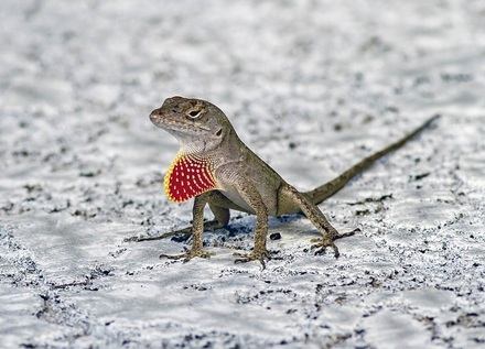
The green anole lizard, or Carolina anole (Anolis carolinensis), is a common laboratory species. Basic rules of its biology—for example, how it responds to exercise—ought to apply to other vertebrates, such as humans. In the past, scientists have successfully used exercise to increase endurance in frogs, birds, alligators and crocodiles. But the same efforts with lizards have been inconclusive.
Jerry Husak, a biologist at the University of St. Thomas in Minnesota, studies lizards with the help of undergraduate researchers. He and his students decided to try creating “Olympic lizards.” They would train their subjects for two kinds of athletic ability, neither of which was totally foreign to the reptiles. Some lizards would become endurance athletes; this long-distance locomotion would mimic the slow patrolling and foraging anoles do in nature. And other lizards would become sprinters; in nature, they use bursts of speed to escape predators.
Thirty lizards were divided into sprinters, distance runners, and a control group. The sprinting track was a dowel two meters long and five centimeters wide, propped at a 45-degree angle. The researchers chased the lizards up the dowel and used infrared beams to measure their fastest speed. Sprinters “trained” three days a week for eight weeks. Gradually, the researchers increased the training intensity by making the lizards do more runs per day.
Meanwhile, the distance runners did their training on a treadmill. The researchers set the treadmill to a low speed and gently prodded the lizards with a paintbrush to keep them moving. These athletes had to stay on the treadmill for 30 minutes at a time, or until they were exhausted. (How do you know anoles are exhausted? “When we flip them over onto their backs and they can no longer flip themselves back onto their feet,” Husak explains. Glad he’s not my trainer.) These lizards, too, exercised three times a week for eight weeks, while the steepness of the treadmill gradually increased.
At the end of the training regimen, the researchers tested all their lizards a final time. The distance runners had clearly improved. On a fast treadmill, the endurance-trained lizards could run for almost three times as long as they had initially. Blood samples showed that their hematocrit levels—a measure of red blood cells, which carry oxygen—had also increased. And dissecting the limbs of dead lizards revealed that their muscle fibers had grown, just as they do in exercising mammals.
The sprinting lizards were a little more disappointing. In their final trials, they didn’t run any faster than they had before training. But their muscle fibers had also grown. Husak suspects that these athletes had actually improved—they just didn’t feel like performing.
“I definitely think the sprint-trained ones increased their sprinting abilities,” Husak says. But after the lizards had spent so much time being handled by humans, he says, “We just couldn’t motivate (i.e., scare) them enough…to run as fast as they could.”
There’s not likely to be a lizard Olympics anytime soon. Creating athletic anoles isn’t the only goal of Husak’s research, though. He’s ultimately interested in the tradeoffs that come with being a good athlete. Animals that spend more energy on reproduction, for example, may have to sacrifice life expectancy or immunity. Do the same tradeoffs happen when animals spend their resources to build beefy muscles?
Husak has gotten closer to answering that question by showing that lizards can be trained. Now he just has to figure out how to scare them into performing their best—because even if the biology of exercise is the same across vertebrates, the power of a “Just Do It” poster isn’t.


