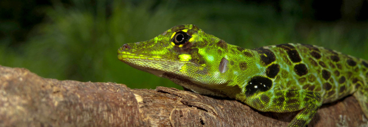
Anolis sagrei Survey Continued: Eleuthera, The Bahamas

beach scrub and bay scenic 2

I just got back from a short trip down to Eleuthera in The Bahamas where I was assisting Anthony Geneva (Harvard post-doc) in sampling lizards. Also along for the trip were Sofia Prado-Irwin (Harvard Ph.D. student) and Rich Glor (University of Kansas). We went with the main goal of sampling Anolis sagrei from four habitat types found commonly in the Bahamas as an extension of an ongoing project in the Losos lab (previous posts from: Rum CayConcepcion IslandRagged IslandBiminiMangrove habitat, and Great Isaac Cay). Specifically, we were looking to sample Anolis sagrei in mangrove, secondary coppice forest, closed coppice forest, and beach scrub habitats. These habitats differ in the height of the canopy, density of the understory, and composition of plants.

This slideshow requires JavaScript.

We focused entirely on the southern half of the island near Rock Sound and Cape Eleuthera. We were successful in sampling two beach scrub habitats, two mature coppice forest, one secondary coppice forest, and one mangrove habitat. We were able to catch all four of the anole species found on Eleuthera: Anolis angusticeps, Anolis distichus, Anolis sagrei, and Anolis smaragdinus. We also encountered a number of other native herp species: the Bahamian boa (Chilobothrus striatus), Ameiva auberi, Eleutherodactylus rogersi, curly tailed lizards (Leiocephalus carinatus), and the Bahamian racer (Alsophis voodoo), as well as a couple of non-native species: Cuban tree frog (Osteopilus septentrionalis), and Hemidactylus mabouia.

This slideshow requires JavaScript.

In my own research I work with Anolis cristatellus, the Puerto Rican crested anole. I am always surprised when I catch A. sagrei by how much smaller they are than A. cristatellus, although very similar in appearance otherwise. On this trip, I was also surprised that the A. sagrei, as well as the A. angusticeps and the A. smaragdinus, appeared to be much smaller than those I had encountered on Bimini last spring.

We also found that the density of lizards was quite low compared to what we expected and what I had experienced in Bimini, both during the day and at night. In all four of the habitat types, we saw an abundance of hatchlings, juveniles, females, and small males, but relatively few full adult male A. sagrei. For A. angusticeps and A. smaragdinus, we encountered only a few individuals total during the week of sampling. This reminded me of an odd experience I had last fall in Puerto Rico with A. cristatellus. It was the same time of year and I had an extremely difficult time locating mature animals in sites where I had previously sampled large numbers during the spring and summer months. Instead, I observed a large number of very young animals and females. I’m curious if this is a coincidence or if perhaps there is a strong seasonal effect on either male behavior (i.e., reduced visibility outside of the mating season) or male abundance (i.e., reduced numbers because of mortality during the mating season). In other words, are the males still there, but hiding, or are they really lower in abundance in the late fall? Or maybe I was coincidentally unlucky on both trips… I am very curious to hear thoughts on this!

Anolis sagrei using coral ground habitat.

Anolis sagrei using coral ground habitat.

Finally, I want to end with a short natural history note on the habitat use of the A. sagrei in the mangrove habitat. In this habitat we observed A. sagrei using perches at drastically different heights: some were 6 feet up, others were on the ground. Interestingly, the ones on the ground did not appear to be in transit, but seemed to be using the pockmarked karst as perches, running into one of the many holes when approached. Has any one else observed this behavior before? It seems so different from the typical trunk-ground anole perch and behavior to me.

That’s all for now. Currently Anthony is sampling additional islands in the Bahamas along with Melissa Kemp (Harvard post-doc) and Colin Donihue (Yale Ph.D. candidate / Harvard visiting student). Best of luck to them, I can’t wait to hear how the rest of the trip went!

Reminder: Submit Photos for Anole Photo Contest 2015!

Steven De Decker and Tess Driessens’ winning photo from the 2012 contest.

Thank you to everyone who has sent in photos for our 2016 calendar contest, we’ve received some great submissions! For those who haven’t yet gotten around to it, you’ve still got one week – the deadline is Saturday, November 21. If you have any great anole photos, we would love to have them in the contest! And just to remind you, the first and second place winners will receive a free Anole Annals 2016 calendar, woo! So send in your pics, let’s make the 2016 calendar great!

To remind you, here are the rules: submit your photos (as many as you’d like) as email attachments to anoleannals@gmail.com. To make sure that your submissions arrive, please send an accompanying email without any attachments to confirm that we’ve received them. Photos must be at least 150 dpi and print to a size of 11 x 17 inches. If you are unsure how to resize your images, the simplest thing to do is to submit the raw image files produced by your digital camera (or if you must, a high quality scan of a printed image).  If you elect to alter your own images, don’t forget that it’s always better to resize than to resample. Images with watermarks or other digital alterations that extend beyond color correction, sharpening and other basic editing will not be accepted. We are not going to deal with formal copyright law and ask only your permission to use your image for the calendar and related content on Anole Annals (more specifically, by submitting your photos, you are agreeing to allow us to use them in the calendar). We, in turn, agree that your images will never be used without attribution and that we will not profit financially from their use (nobody is going to make any money from the sale of these calendars because they’ll be available directly from the vendor).

Please provide a short description of the photo that includes: (1) the species name, (2) the location where the photo was taken, and (3) any other relevant information. Twelve winning photos will be selected by readers of Anole Annals from a set of 28 finalists chosen by the editors of Anole Annals.  The grand prize winning and runner-up photos will be chosen by a panel of anole photography experts. Deadline for submission is November 21, 2015.

Good luck, and we look forward to seeing your submissions!

Fixing Formalin: Perhaps Not a Game-Changer, But a Good Start!

Sarah Hykin and Jim McGuire

In our recently published paper in PLoS One, we provided ‘proof-of-concept’ that it is possible to obtain genome-scale data from formalin-fixed specimens. This study was proposed by one of us (SMH) about four years ago while contemplating how she might be able to undertake a phylogeographic study of a rare lizard species for which there was little hope of resampling the entire range.

Of course, people have been contemplating the challenge of obtaining DNA sequence data from museum specimens for decades, often with limited success. The typical approach involved targeting mitochondrial genes, developing sets of nested primers amplifying short fragments (often only 50-100 bp in length), and then a brute-force amplification and sequencing in an effort to score a few hundred usable base pairs.

However, our discussion was informed by the recent development of short-read Next-Generation Sequencing on the Illumina platform, which produces genomic-scale data 50-100 bp at a time. Surely, we thought, if any method could efficiently pull DNA sequence data from formalin- damaged DNA, this was it. Our timing was impeccable because our campus had just obtained a major foundation grant to support, among other things, the development of risky technology that could enhance the utility of historical museum specimens. We obtained a small subaward and Sarah went to work studying the literature on historical DNA sequencing, and figuring out how to perform NGS. Illumina sequencing and bioinformatic processing have become pretty routine now, but this was a very challenging undertaking at the time, and Sarah had to pursue this while focusing her energies on her unrelated dissertation research.

The first decision that we had to make was which species and specimens to select for sequencing. This turned out to be a ‘no-brainer’ because the only squamate genome available was Anolis carolinensis and we needed a genome to which we could map reads. Being a conservative museum curator, Jim suggested using no-data or limited-data specimens so that when the project inevitably failed we would not have cut up particularly important specimens. In retrospect, this was a mistake. For example, we used a limited-data specimen of Anolis carolinensis that was in the database as having been obtained in 1985. Further investigation of this specimen would have shown that this date was dubious, and it now appears the specimen was actually obtained and prepared between late 1986 and 1988 (and accessioned in 1990). We also have reason to believe the specimen was fixed in buffered formalin for one-week prior to rinsing and immersion in ethanol, though this is not known for sure since the specimen does not have associated field notes. If we had this to do over again, we would make sure that we knew as much as possible about the source specimens rather than taking a ‘let’s limit the damage if this experiment fails’ approach to specimen selection.

It works (sometimes)!

As indicated in our paper, it is indeed possible to obtain genome-scale data from formalin-fixed specimens housed for decades in a museum collection. However, our method is far from fool-proof and we strongly suspect that idiosyncratic features of individual specimens will determine success or failure in many instances. The age of the specimen is likely to be one of the most important variables as suggested by our published study – sequencing of our 100-year old sample failed, whereas sequencing for our ~25 year old sample was successful. However, other parameters are likely to prove important. For example, others have shown via direct experiments that DNA is better protected by buffered (versus unbuffered) formalin. We also suspect that other features of the specimen’s preparation such as the time spent soaking in formalin prior to immersion in ethanol, the concentration of the formalin used, the quantity of formalin injected into the specimen, and the time that passed between the death of the specimen and its preparation could all make a difference. These conditions are rarely recorded at the time of preparation, which means that for the vast majority of specimens that might be targeted for NGS, the researcher cannot know ahead of time whether the specimen is likely to be a good versus poor candidate for sequencing.

Our paper received some attention on social media with some calling our study a “game changer” and others arguing that such a statement is overblown. From our perspective, this is semantics. Have we completely solved the issue of obtaining genomic sequence data from formalin-fixed samples? Certainly not. Have we identified in a controlled way, the precise conditions underpinning success or failure of NGS from formalin-fixed samples? Again, not by a long-shot.

However, we have shown some things that are likely to be important in moving this technology forward (which could be interpreted as changing the game). Most importantly, we have shown definitively that it is possible to obtain genomic data from old museum specimens. This had not been shown previously and we believe that this will encourage many more people to give it a try than would be the case if our paper had appeared in the Journal of Negative Results. Further, we were able to shed some light on methodological issues that are actually quite important. First, we obtained a sufficient quantity and quality of DNA for sequencing from liver tissue and not from either leg muscle or, most importantly, bone. Many of you will be aware of a terrific study published by Maureen Kearney and Bryan Stuart in 2004, in which they provided a phylogeny for amphisbaenians that was based in large part on sequences obtained from old museum specimens. In their groundbreaking study, they obtained mitochondrial and nuclear sequence data using laborious traditional Sanger sequencing of short DNA fragments, along with non-traditional extraction of DNA from bone tissues using methods developed for human forensic DNA analysis. Their successful extractions required sampling bone from pickled specimens, which can be quite destructive. In contrast, pulling liver from a museum specimen is minimally invasive – especially when you consider that most newly collected specimens will have had their livers removed prior to preparation. Thus, our finding that liver is an optimal DNA source for NGS is important.

Further, second, we found that a modified phenol-chloroform extraction protocol outperformed Qiagen extraction for NGS purposes. Indeed, we now suspect that our failed attempt to perform NGS on our older sample could very well be the result of our effort to systematically compare extraction protocols. For both of our Anolis specimens, we subsampled the entire liver, which was divided into a small piece and a much larger piece. For the younger sample, the larger piece was extracted using phenol-chloroform, whereas for the older sample, the larger chunk was extracted using Qiagen. Importantly, from that older sample,we obtained more DNA from a sample ~20 times smaller in mass using phenol-chloroform versus Qiagen extraction. If we had dedicated the larger subsample from the 100-year old specimen to phenol-chloroform, we believe this might have resulted in successful NGS.

Fig4

Figure: Sample placement in the phylogeny. As expected, our sequencing effort generated low-coverage (~0.5X) of the Anolis carolinensis genome. However, we did obtain ~60X coverage of the mitochondrial genome, providing a means of evaluating the quality of our sequence data after processing. We aligned the ND2 sequence from our sample with GenBank sequences representing A. carolinensis from Louisiana, the Anolis carolinensis complete genome, a variety of additional Anolis species, and a more distant outgroup. The phylogram shown here suggests that our mitochondrial sequence data obtained from a formalin-fixed specimen are reliable. If sequencing errors were evident, we would minimally expect the branch representing our formalin-fixed specimen to be relatively long compared with other Louisiana A. carolinensis, or perhaps even misplaced on the tree.

Where to from here?

One critique that we received in review was that we had failed to perform rigorous controlled experiments testing the various conditions that could impact success with NGS from formalin-fixed samples. Though we would have loved to perform such experiments ourselves, even limited NGS sequencing is still sufficiently expensive (thousands of dollars, rather than hundreds) that we were not in a position to pursue this. However, we would love to see someone – perhaps reviewer number 2 – grab the bull by the horns and perform this experiment! Such a follow-up study, should it identify via controlled experimental procedures the key parameters for successful NGS of formalin-fixed samples, would come closer to meeting the criteria of being a ‘game-changer.’

Blanchard Cave, a Window into the Late Pleistocene and Holocene Squamates from Marie-Galante Island (Guadeloupe Archipelago, Lesser Antilles)

Over the past few years, two European research programs developed an interest in the ancient fauna and environment of the Guadeloupe islands. The prospection for cave deposits led to the discovery of numerous accumulations of fossil remains documenting the Holocene and Late Pleistocene faunas of the archipelago, especially on the island of Marie-Galante, where three major deposits were discovered.

Blanchard Cave is one of these deposits. This cave contains the oldest fossil-bearing sedimentary layers of the island dated around 40,000 years before present and is an excellent complement to the two others cave documenting the Late Pleistocene fauna of Marie-Galante (Cadet 2 and Cadet 3).

After a test excavation in 2008 that revealed the potential of the site in term of fossil fauna, Blanchard cave was investigated between 2013 and 2014 in the framework of a European research program interested in the past environment and fauna of the Guadeloupe islands, the BIVAAG project. The three excavation campaigns conducted during this period allowed the precise documentation of the sedimentary filling of the cavities and the recovering of thousands of skeletal remains mainly attributed to frogs, lizards, snakes and bats.

The excavation work in the cave (Picture: A. Lenoble)

The excavation work in the cave (Picture: A. Lenoble)


Welcome gifts from the bats… (Picture: C. Bochaton)

Welcome gifts from the bats… (Picture: C. Bochaton)

But collecting the fossils remains was not that easy and although the perspective of working in the Caribbean a few hundred meters from the sea could seem very attractive, the working conditions in the cave were far from pleasant. Mainly because the cave was inhabited from the ground to the roof by numerous cockroaches, rats, gnats and bats. Bats were extremely noisy, and proved to be extremely rude hosts. Another difficulty was the potential occurrence of histoplasmosis in the cave that led to the necessity of wearing a respirator during the work. Such masks make breathing difficult during the work and combined with the heat, humidity and other disagreements previously mentioned strongly impact your initial enthusiasm.

Once you overlook these difficulties, the sediment was extracted from the site and then washed and sieved in order to retrieve the small bones contained in it (the bones are usually smaller than 5 mm). The remains were then recovered and sorted, partly in the field (unfortunately this activity often kept the paleontologists outside of the cave and away from the bats), before being studied.

Washing and sieving of the sediments (Picture: M. E. Kemp)

Washing and sieving of the sediments (Picture: M. E. Kemp)

Recovering of the fossil bones (Picture: M. E. Kemp)

Recovering of the fossil bones (Picture: M. E. Kemp)


The results of the study of the squamates remains collected in the cave can be found in a very recently published paper. To summarize the main findings, we found evidence of the past occurrence of at least ten species of snakes and lizards: four snakes: Antillotyphlops sp., Boa sp., Alsophis cf. antillensis and an undetermined colubroid; and six lizards: Anolis ferreus, Iguana sp., Leiocephalus sp, Thecadactylus sp., cf. Capitellum mariagalantae and Ameiva sp.. The stratigraphic distribution of these taxa in the site combined with previously existing data show that only two extinctions (Boa sp. and Colubroid ind.) are dated from the Pleistocene/Holocene transition and thus predate the arrival of humans on the islands around 5000 years ago. Then during the pre-Columbian times two new taxa appear in the deposits, Iguana and Thecadactylus. On the other hand, a massive faunal turnover began after the European colonization of the island. Indeed, at least six squamate genera (Leiocephalus, Capitellum, Ameiva, Antillotyphlops, Alsophis and Erythrolamprus), including all the snake genera, were extirpated between 1492 and today. Thus, 55% of the squamate genera present during pre-Columbian times went extinct over the past few centuries.

These results are further evidence of the current sixth mass extinction crisis and of the strong impact of humans on this insular fauna. However, Marie-Galante Island remains an isolated case because the past fauna of most of the Lesser Antillean islands remains poorly known and in most cases totally unknown despite the critical importance that such data may have in many fields to test inferences built on modern data.


To Eat or Be Eaten: How an Anole Decides When to Forage

Anolis cristatellus in survey posture (photo by K. Winchell)

Anolis cristatellus in survey posture (photo by K. Winchell)

Foraging decisions are the result of a complex decision-making process involving intrinsic factors (physiology, body condition, cognitive ability, sex, ontogeny, etc.) and environmental factors (food availability, structural habitat, presence of predators and competitors). In short, it comes down to the tradeoff between the benefits of energetic gain and the potential costs of predation risk, missed opportunities for reproduction, and expended energy. However, little is known about the specifics of this process – what information are lizards considering when making this decision? By conducting manipulative field experiments on Anolis cristatellus in Puerto Rico, Drakeley et al. (2015) attempt to elucidate what environmental factors influence the decision to forage.

The authors conducted field experiments involving feeding trays in the wild. The Puerto Rican crested anole is a trunk-ground anole and a sit-and-wait forager. When receptive to feeding, it perches head down in “survey posture,” a behavior it reduces when satiated. Aside from movement associated with foraging and social interactions, this species typically remains stationary on a perch. Because of this, the authors were able to easily locate a focal individual and count the number of conspecifics present, using natural variation instead of manipulating the number of animals present.

In the first experiment, they manipulated the food quantity to determine how foraging decisions differ when food is plentiful versus scarce and how this is influenced by the presence of competitors. They found that lizards foraged faster when there were more conspecifics present and food was scarce. When no lizards were near the feeding tray and the feeding tray was full, the focal animal took longer to approach the tray to take the mealworms compared to when there were many conspecifics nearby. Interestingly, this was not related to overall local density, but rather to the number of conspecifics in the immediate vicinity. Therefore the decision to forage likely involves an instantaneous assessment of the local conditions rather than knowledge of the long-term population trends. The authors also considered several other factors and found that although body size was related to foraging latency (larger lizards were quicker to the feeding tray), no other environmental factors were relevant (temperature, humidity, perch height, perch diameter, local density of conspecifics).

Figure 1 from Drakeley et al. (2015). Latency to feed was correlated with the number of conspecifics present and abundance of food.

Figure 1 from Drakeley et al. (2015). Latency to feed was correlated with the number of conspecifics present and abundance of food.

In the second experiment, the authors chose focal animals farther from the feeding trays and considered distance as a proxy for predation risk. The farther the lizard was from the tray, presumably the greater exposure it had to predators as it moved towards the tray. They found that under this scenario, when risk was elevated, there was more latency in the approach of the food tray. This effect was driven mainly by the increased use of intermediate perches rather than a direct approach across open ground. Increased latency to feed was observed regardless of how abundant the food was or how many conspecifics approached the tray, supporting the conclusion that this effect was because of the perception of greater predation risk (i.e. movement over a longer distance). They also found that larger lizards had a lower latency to feed (approached the feeding tray more rapidly) and lizards not in the foraging position had a longer latency to feed.

In summary, it seems that anole foraging decisions are quite complex. Lizards appear to weigh the risk of predation taking cues from conspecific behavior and abundance versus the abundance of food to make instantaneous decisions to approach a novel feeding source.


Drakeley M, Lapiedra O, Kolbe JJ (2015) Predation Risk Perception, Food Density and Conspecific Cues Shape Foraging Decisions in a Tropical Lizard. PLoS ONE 10(9): e0138016. doi:10.1371/journal.pone.0138016

Reaching Safety

When somebody talks about roads crossing along natural forest, we could think about the perturbation this may cause to local fauna, especially in the Tropics. At least in Panama, wildlife crossings are not so popular in terms of design, deployment and monitoring. To my knowledge, the few existing ones are aerial and designed keeping in mind the crossing of monkeys or sloths for example. This issue came to my mind on the 3rd of November when I saw a Dactyloa insignis trying to cross an 8 m road traversing Santa Fe National Park, one of the pristine forest in central Panama.

Captured at Santa Fe National Park, Panama

Captured at Santa Fe National Park, Panama

It made three short attempts and looked clumsy when trying to run on the pavement puting him at risk of death, so we caught him and helped him reach the other side of the road.

Should the Use of Subspecies Be Revived?

The last week has seen a spirited discussion of the pros and cons of splitting recognized genera into multiple, smaller genera. We’ve had 34 comments already. Check it out! And if you’re an advocate of splitting genera, that viewpoint has been getting the short end of the stick and could use more support.

As a tangent, the topic of subspecies has come up, and David Hillis has strongly argued for reviving its use. Here’s what he has to say:

First, I don’t think either species or subspecies are “clades.” Species are lineages (the branches on the tree of life). Sexual recombination among individuals results in tokogenetic relationships within species. Clades, on the other hand, are monophyletic groups of lineages on the tree of life. Rather than being defined by tokogenetic relationships, they are defined by phylogenetic relationships.

Traditionally, subspecies are geographical races of species. In other words, they are geographically distinct populations that nonetheless meet and interbreed at contact zones. Sometimes, these contact zones are very broad, as with broad-banded versus southern copperheads. If the contact zones are very narrow, and there is strong evidence that the contact zone is a genetic sink (there is no gene flow across the zone, because of strong selection against hybrids), then I agree that the two entities can be considered separate lineages, and hence species. But in many recent cases, as with the copperhead example, there is abundant evidence that the contact zone is NOT a sink, and that there is NO selection against hybrids. In this case, I disagree strongly with the authors who proposed to split these subspecies into distinct species. That is inconsistent with any lineage species concept…there is a huge area where these two forms intergrade, with no evidence of any loss of fitness. Thus, the two forms are geographical, intergrading races, or subspecies.

I think we will soon see a backlash against the splitting off of geographic races as species as well. Frank Burbrink (who was an author on the copperhead example I mentioned above) and I plan to write a pro/con article about this together, each arguing our respective points of view. Hopefully, this will re-kindle the conversation about subspecies.

Subspecies are unpopular right now because they were long abused in several ways. Inappropriate uses include (1) to describe non-geographic “varieties”; (2) to arbitrarily break up clines; and (3) to describe distinct, isolated lineages that clearly are species. But used in proper context to designate a geographically distinct race, they are certainly reasonable and often useful. They are rarely used in some groups, for several reasons: Groups like freshwater fishes have discrete ranges, so taxa don’t interbreed over broad areas. And many groups are too poorly studied to understand geographic variation. But in well-studied terrestrial groups (like herps), subspecies are perfectly reasonable and useful taxa to designate intergrading geographic races.

Dragons from the Old World

The Neotropical and Oriental realms both were once a part of Gondwanaland. Interestingly, both of these realms exhibit same ‘type’ of lineages occupying equivalent niches. Boas dominate the Neotropical zone whereas pythons flourish in the Oriental. Similarly, in the Old World (or Oriental or Indo-Malayan realm), there are lizards belonging to family Agamidae which exhibit uncanny parallels to Anolis sp. in their natural history.

CSC_5284One example is from Yelagiri Hills in the Eastern Ghats region of the Indian state of Tamil Nadu. This is Psammophilus dorsalisDuring the breeding season, males of this species turn their drab and dull dorsal region to bright yellow or red to impress conspecific females. The brighter the male, the more chance he has to win over females. Males display such behavior for the entire day; at night these lizards hide under rocks.

When equally bright males encounter each other, competition is settled by ‘ducking’ heads and throwing off the opponent from the rock.

Taxonomic Splitting Revisited: When Should Genera Be Subdivided?

Over the last several years, ever since Nicholson et al. proposed dividing Anolis into eight genera, the topic of taxonomic splitting has periodically been discussed in these pages (for example, this post, its comments, and links to other posts).

The general question of when to split taxa recently has been revisited in several comments in AA. A week ago, David Hillis wrote:

Anolis is a valid name for a monophyletic group on the Tree of Life. It is “special” as a genus only in that the genus name is used as part of a binomial for particular species. It doesn’t make sense to change the scope and application of generic names unless the names are actually misleading about phylogeny (e.g., if Anolis were polyphyletic, then that problem should be fixed). But splitting a valid, monophyletic genus into a bunch of smaller genera, and thereby needlessly changing the names of many species, without fixing any phylogenetic problems with the existing taxon names, is not science. It is just playing around with names. If someone wants to name the groups within genera, then do so…but there is no reason to change the meaning of a existing name (or the names of the all the affected species) in doing so. That is the kind of silliness that gives taxonomists such a deservedly bad reputation among biologists.”

Elswhere, David posted a flowchart on his recommended decision-making process about whether and how to divide recognized genera:

Hillis flow chart

Ivan Prates, in line with comments he made in a recent paper on A. punctatus, then remarked:

“This seems more like a sociological matter.

During the ‘taxonomic revolution’ of the amphibians, about 10 years ago, the (perhaps?) most influential (or faster?) group was the splitter one, and their taxonomic scheme prevailed. Currently, nobody is upset about which species were once named as Bufo, Hyla or Rana. A few do care about Dendrobates – like Anolis, a sexy group with a body of dedicated investigators.

It seems that a single genus makes sense for the community that investigates dactyloid lizards more closely. On the other hand, those who deal with overwhelming levels of herpetological diversity in the tropics (waaaay beyond lizards) see benefit in more partitioned schemes, which correlate more closely to morphology and geography.

So, when we discuss names, it may be healthy not to forget about our diversity as investigators as well. About science, splitting Anolis is not science, but well, not splitting Anolis isn’t science either.”

On the Origin and Diversification of the (Hemi)Penis: Anolis Takes Center Stage

Over the last decade the term “model species” has taken on new meaning. Species that were once the building blocks for distinct disciplines have taken on new importance in comparative evolutionary studies that integrate perspectives across biological disciplines. Nowhere is this better illustrated than with Anolis lizards. For decades anoles were a workhorse of ecologists and evolutionary biologists, but have, more recently, been embraced by developmental biologists, genomicists, physiologists, and neurobiologists among others. This disciplinary expansion is perhaps most evident with the rapid increase of penis/hemipenis research that has been published using anoles within the year.

For many herpetologists, including those focused on anoles, the hemipenis is ripe with taxonomic characters, easily allowing for the identification of new species. Julia Klaczko and colleagues recently demonstrated that features of the hemipenis are some of the most rapidly evolving characters among anoles, a group already well known for its rapid anatomical evolution. Independent from these taxon-specific interests, developmental biologists became interested in the anole hemipenis because of its unique anatomy compared to other amniotes. Marissa Gredler and members of the Cohn Lab used anoles as one of their reptilian models of external genital development in what is arguably the broadest embryological survey of reptilian phallus development to date. In parallel, Patrick Tschopp and colleagues probed the cellular and molecular regulation of early phallus development among anoles, snakes, chickens and mice, demonstrating that the hemiphalluses (hemipenes and hemiclitores) and hindlimbs of squamates utilize similar molecular networks at the earliest embryonic stages of morphogenesis. Now, just within the last month, two more papers have used anoles in studies of phallus evolution and development, one using cutting-edge molecular techniques to better understand the relationship between limbs and external genitalia and the other addressing the fundamental question of external genital homology using museum specimens that are more than 100 years old.

Before getting into the findings of this new research, lets lay out some of the dirty details of penis evolution. First and foremost, the penises of amniotes are extremely diverse. Squamates have paired lateral phalluses while other clades have a single midline phallus. Each of the amniote lineages uses hydrostatic pressure to achieve an erection, yet accomplish this using different bodily fluids (lymph or blood). In mammals sperm is transferred to the female through a closed urethral tube, but other groups utilize an open channel. Most birds (97%) and the tuatara, have absent or highly reduced phalluses and reproduce with the famed “cloacal kiss.” These large differences in anatomy should not overshadow the spines, bulges, corkscrews, and dramatic differences in size that give species their distinctive features. But with such striking variation, we are forced to wonder how many times the penis evolved. Perhaps the amniote ancestor possessed an intromittent phallus capable to transferring sperm to the female that later diversified in each lineage independently. Or, perhaps the last common amniote ancestor used cloacal apposition to foster internal fertilization and unique phallus morphologies evolved independently at the origin of each lineage. Because adult anatomy provides few clues to phallus homology, Thom Sanger (me), Marissa Gredler, and Marty Cohn looked towards the embryo for help.

Table 1 from Sanger et al. 2015 summarizing phallus variation in amniotes

Table 1 from Sanger et al. 2015 summarizing phallus variation in amniotes

The tuatara, a species lacking an adult phallus, has presented a problem in attempts to reconstruct the last common ancestor of amniotes because it raises the distinct possibility that reproduction through cloacal apposition was the ancestral condition.

Page 116 of 297

Powered by WordPress & Theme by Anders Norén