
The dewlap is probably the most noticeable thing about anoles. For me, the best way to spot an anole is by the flash of color from the dewlap as a lizard displays. Without that, many anoles would remain cryptic amongst the vegetation. This seems to be the case for the lizards themselves as well. The burst of color and movement as the dewlap is rapidly extended is a wonderful device for attracting the attention of rivals and mates. It’s possible that the dewlap originally evolved as an attention-grabbing flag to augment an existing sequence of elaborate headbob movements in forested environments. These days, the dewlap is a complex signal component in its own right, often with a dizzying array of colours and displayed using a variety of movements.
Anoles aren’t the only ones with a moveable dewlap. The Southeast Asian Draco lizards have a dewlap, and again to back up the headbob movements that make up their main channel of social communication. There are many other parallels between Draco and Anolis lizards, but the similarities in how they communicate is something that particularly fascinates me.
Early on in my fieldwork with Draco, I started discovering species that didn’t seem to use headbobs as part of their social display. It seemed these species had lost the headbob entirely and instead concentrated all of their communication through the dewlap display. These species are a minority, but not by much. It was a puzzle. These Draco had lost a central and complex element of their communication in favour of something that was seemingly more basic. Communication biologists are often fixated on trying to explain how animal communication becomes more elaborate over evolutionary time, but less attentive to why complexity subsequently becomes lost. These Draco lizards were an excellent case study.

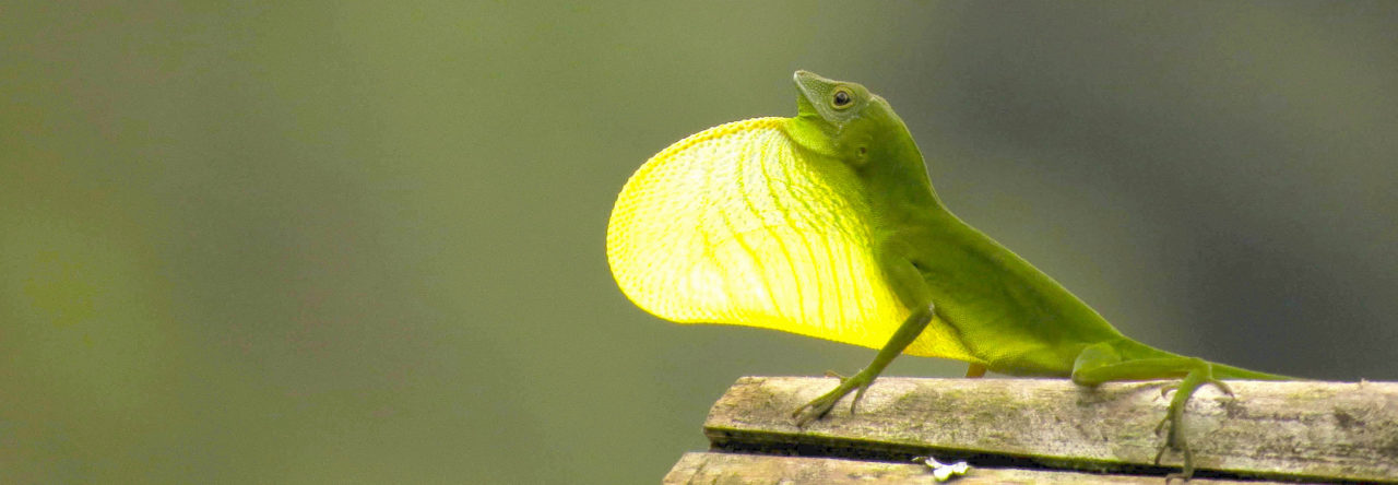
Draco melanopogon (photo above) only communicates using the dewlap, whereas Draco sumatranus (opening banner photo) relies on both headbobs and the dewlap, just like anoles.
After nearly a decade of fieldwork on numerous species of Draco throughout Malaysia, Borneo and the Philippines, my trips stalled in 2020, as did the rest of the world. Celebrities had nothing better to do than write biographies, but my lockdown project was to focus on using the data I already had at hand to finally solve the curious case of the missing headbob.
It felt like an endless series of stay-at-home orders in Australia, and well into 2021 too. While the celebrities had gone on to finish their books and were now doing the zoom promotion circuit, my progress was hurdled by home-schooling two young children. We survived home-schooling in the end, and my attempt at figuring out why some Draco have lost the headbob has finally been published.

The evolutionary history of visual displays in agamid lizards
The first discovery is the headbob display is very ancient, evolving something like 130 million years ago or more. That’s before the evolution of Draco, and before the evolution of the anoles, in an evolutionary ancestor to both the iguanid (new world) and agamid (old world) lizard families. This was back in the age of the dinosaurs. Today, virtually all iguanid and agamid lizards use a headbob display or some variant of it in social communication. Which means the absence of the headbob in a handful of Draco species is very unusual.
The loss of the headbob from the social display of Draco is effectively a loss of complexity. A loss of complexity means a loss of “information potential.” Try writing a biography with half the alphabet. You might manage the following or something a little longer: “I was born. I paid taxes.” Thirteen unique letters in total. Obviously not the rich backstory you might hope for. Not because you hadn’t lived a fulfilling existence; rather you don’t have the language complexity to convey it in detail.
There are various reasons animals might lose complexity in their social signals. Perhaps the original need for a complex signal is no longer present. Perhaps the invasion of a new environment puts a brake on the level of complexity that can be accurately perceived. Or perhaps natural selection on other things, like body size, has made performing a complex signal too costly.
The beauty of having spent so much time in the field is the accumulation of a large library of data. By leveraging this information, I was able to test each of the above scenarios. The short of it is, Draco that have lost the headbob are unusually large species. Physically moving the head and body in a headbob display is more energetically expensive than pumping the dewlap in and out. It seems, then, that the physiological cost of performing the headbob became too great for these large species and they shifted to relying only on the dewlap for communication. This implies the communication system of these species is compromised, unless they have made up the loss of information potential somewhere else.

Draco without the headbob have more complex dewlap colour patterns. Each dot is a different species.
In fact, the dewlap itself tends to be more complex in Draco that have lost the headbob. Stealing a method for measuring complexity of anole displays, the dewlap of these Draco are more elaborately coloured than the average Draco. Unfortunately, this is unlikely to have been enough to fully cover the loss of the headbob. This means Draco that no longer use the headbob are relying on a constrained communication system.
The idea that the headbob is likely to be more energetically expensive than the dewlap was originally proposed for the anoles. It was used to explain the physiological basis for why Jamaican anoles might have evolved an innovation that allowed them to move away from a headbob-centred display in favour of one focussed on the dewlap. To be clear, the Jamaican anoles do still rely on headbobs in their social displays. But a rapid series of dewlap pumps features more prominently in their displays compared to the typical anole, like those on Puerto Rico for example.
It seems the dewlap has begun to supersede the headbob in anoles as well.
If you’d rather not slog through the paper itself, you can view a 12 minute video summary instead. If you would like to slog through the paper and can’t access it behind the paywall, drop me an email and I’ll forward you a free copy (t.ord@unsw.edu.au).
 English translation


 While hunting sagrei for fun this evening with my 3-year-old, we spotted a large male Anolis sagrei eating what I think is a mourning gecko (Lepidodactylus lugubris). This happened at 7:18 pm in the presence of minimal artificial light from lamps (albeit strong artificial light from my headlamp). Plenty of sagrei were already asleep on leaves. Surprising to find a diurnal lizard preying on a nocturnal/metaturnal gecko.


