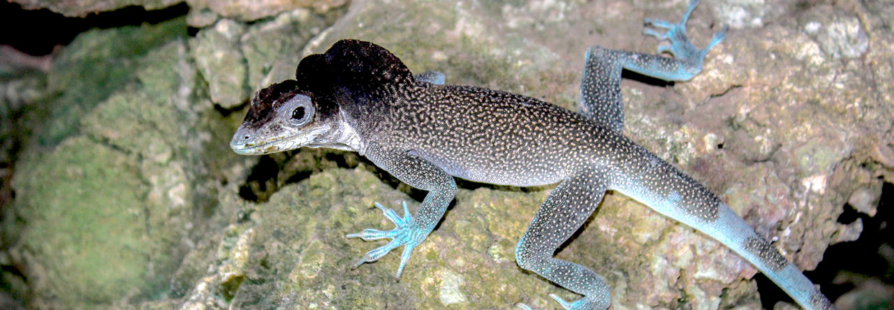
Category: All Posts Page 34 of 145

Seeking Support for New Research Investigating Color Change in Green Anoles

Victoria Pagano’s page from the crowd-funding platform Experiment

Green anoles (Anolis carolinensis) are talked about quite frequently here on Anole Annals, with 11 articles being published in 2018 and 2019 combined! As I am sure many of you are aware, green anoles change color from green to brown, and while it is known how, it is not yet known why. Although there have been multiple field studies into what causes green anoles to change color, the data have been inconclusive. This is why an experimental study is necessary to try to determine the cause of the color change.

In this experimental study, there will be two main hypotheses tested:

The first is the well known thermoregulation hypothesis. I will be testing this by establishing separate light and heat sources, and turning them on and off for different scenarios. If anoles change color for thermoregulation, then they would turn brown more frequently when the heat is off and the light is on.

The second hypothesis is the effect of increased stress. Stress will be induced by sliding a red disk towards the anoles multiple times at a high speed. Any color change that occurs within the red disk moving and the following 10 minutes will be documented as stress-induced.

I will not be able to test the advertisement signaling hypothesis due to feasibility. Because funding and space is limited, I do not have the capacity to house male anoles, as each one needs his own setup. Therefore, testing only females is the only feasible option, and by doing so, the advertisement signaling hypothesis will not be able to be tested, as this hypothesis pertains mainly to males.

To raise funding for this project, I am using an all or nothing crowdfunding platform called Experiment. As fellow anole lovers, I hope that you can help support my scientific endeavors by visiting my project page. All forms of support are greatly appreciated, from donations, to telling your friends about the project, or even by just reading my project page and commenting your thoughts! Whatever the contribution, I am very grateful, and am simply excited to be able to share what I am doing with all of you!

If you wish to learn more about this project, you can visit the project page, “What drives the color change in green anoles?”, where I have posted my methodology, protocols, and will be posting continuous updates on the progression of the project. If you become a contributor, you will have exclusive access to more updates, and will be able to learn more about the research.

My project page stops accepting donations on November 1st at 12:00 AM PT, so be sure to make your way over to the page by then to give your support!

Thank you for taking the time to read this article. I hope that you will explore the project page, and help support this cool and unique research!

Colour Change in the Gorgetal Scales of an Anole Dewlap

An adult male Anolis amplisquamosus with black gorgetal scales immediately after capture (left); the same individual ~10 min later with white gorgetal scales. Photo Credit – John David Curlis


Anole dewlaps are excellent examples of a “complex signalling system.” They exhibit a staggering diversity of colours and patterns. Each dewlap is species specific and adapted to enable these lizards to communicate, attract mates and guard their territories from rivals or competitors. Generally, the colour of a dewlap (and its gorgetal scales) is considered an unchangeable descriptive trait. This colouration is not only relied upon by scientists looking to identify a species, but also by anoles that co-occur and partition with different species in their select niche.

Therefore, it might be surprising to learn that recent observations prove rapid colour change in anole gorgetal scales is possible. The question is, what implications does this have?

A recent publication in IRCF Reptiles & Amphibians details an observation of Anolis amplisquamosus whereby a male individual upon capture possessed black gorgetal scales that quickly changed to pale yellow. Upon consulting the literature, it seems only one prior documentation of colour change in gorgetal scales was reported (Leenders and Watkins-Colwell, 2003), coincidentally also involving a member of the same species clade.

This recent observation of chromatophoric regulation in anole gorgetal scales may be significant in the wider context of anole biology, in confirming photographically that coloration is not always a fixed descriptive or diagnostic feature — at least among members of the A. crassulus species group. Accordingly, this information suggests that some anoles may have the ability to regulate the colour of their gorgetal scales in the same manner as they regulate dorsal and lateral scale colour.

Because the colour of gorgetal scales is a character often used in species identification, understanding the mechanics and the purpose of such a change is crucial; as well as any implications to display behaviour, communication and anole interactions.

Brown Anoles in Montgomery County, Texas

I recently received an email from Chris McMartin, the director of the Southwestern Center for Herpetological Research, about a population of brown anoles near his home in Montgomery County, Texas, just north of Houston. Chris has done a lot of preliminary research to understand how the Montgomery population is spreading, and would like to know how these lizards are related to the larger population in Harris County.

Interested? Keep reading!

With Chris’ permission, I’ve copied part of his email below:

“I’ve been casually (in my free time, mostly in the summer) researching Brown Anoles (Anolis sagrei) and their spread in southern Montgomery County where I live.  As I amass observational data, I’ve noticed the lizards are abundant in some yards/neighborhoods, but nonexistent in adjacent yards/neighborhoods.  I’m slowly trying to piece together additional factors (presence of outdoor cats, prevalence of certain landscaping features including decorative rocks and tropical plants, age of house/neighborhood, use of pesticides, etc.) which may explain not only the disparity in abundance but provide clues as to how to control their expansion.

One big question I have is whether the lizards are naturally expanding from a single introduction long ago (e.g. rapidly moving northward from Harris County, where they occur in densities many times higher than the highest I’ve observed in Montgomery County), or are an amalgamation of numerous discrete introductions (e.g. when a home installs new tropical plants from a nursery/home improvement store).  Brown Anoles first showed up in my yard a little over a year ago, marking an expansion northward of about ¾ of a mile from my previous northernmost observations the year prior.

I have corresponded with Dr. Benson Morrill, who owns Rare Genetics Inc. offering DNA analysis for (at this time) colubrid snakes (primarily for sex determination) and inquired as to the possibility of sending him samples from various neighborhoods in my area in an effort to determine whether they represent a contiguous related population or are the result of discrete introductions.  He says the process to conduct this analysis would be cost-prohibitive for a private individual such as myself, but perhaps a university student would like to take on the project.

As it is, I currently spend what surprisingly-little free time I have in the summer exploring neighborhoods in my neck of the woods and documenting my obervations—around 60 hours this past summer between field work and analysis—and I’m approaching my limit of resources in time (and definitely money, if considering DNA analysis as part of the project). This is where I think perhaps a graduate student might be interested in taking on a study of Brown Anoles as a thesis project…lots of possible threads to pull (competition with natives, rate of range expansion, effect of occasional hard freezes on population, etc.).

I’ve published articles in local magazines/newspapers about the lizards and have a public-opinion survey from a year ago (still awaiting analysis) trying to find any links between various conditions (age of neighborhood, presence of outdoor cats, etc.) and occurrence/prevalence of browns, especially with respect to A. carolinensis.  Some interesting things seem to be occurring.  Anecdotally, browns are eating greens (hatchlings), Broad-headed Skinks are eating browns, and greens are eating the skinks (with photographic evidence)!  Sort of a three-way lacertilian arms race.”

If this sounds like just the opportunity you’ve been looking for, contact Chris at yall [at] mcmartinville.com.

Green Anole Displays at Brown Anole

Photo from Daffodil’s Photo Blog.

Read all about it in the latest post from Daffodil’s Photo Blog.

Daffodil’s Anoles Are Back!

After a bit of a hiatus, Daffodil’s Photo Blog is back with its splendid anole photos and natural history tidbits. Check out the recent offering.

How the Gene Edited Anolis Lizard Came to Be

From the pages of Nova.

BY KATHERINE J. WU TUESDAY, AUGUST 27, 2019 

209376 copy.jpg
This pale little lizard is one of the world’s first genetically edited non-avian reptiles. Image Credit: Courtesy of Ashley Rasys, University of Georgia

Compared to mammals, reptiles have a weird way of reproducing—and in the spring of 2017, that put Ashley Rasys in something of a pickle.

For months, the University of Georgia biologist was struggling to come up with a way to tinker with the genes of the brown anole (Anolis sagrei), a petite, pointy-faced lizard native to Cuba and the Bahamas.

The reptile had initially caught Rasys’ eye because of, well, its eyes. People with albinism often have poor vision due to problems with their foveae, the dense pits of cells at the back of the eyes that confer visual acuity. While foveae are lacking in most mammals, they’re present in lizards—making them intriguing candidates for studying the genes that impact foveal function.

There was just one problem: Reptiles aren’t easy to genetically manipulate. In other common laboratory animals, like mice and zebrafish, a tool called CRISPR has made DNA editing a breeze. The procedure typically involves injecting freshly fertilized eggs with gene-editing machinery, creating a change that would propagate when the cell divided.

But a few quirks ruled out that particular strategy in these lizards. Female anoles can store sperm for many months before fertilizing their eggs internally, making it difficult to time the introduction of the CRISPR cocktail. Anole fertilization also cues the formation of a soft, delicate eggshell that’s hard to penetrate without damaging the embryo.

That meant Rasys and her advisor, Doug Menke, had to get creative. So they decided to shift the injection back a developmental step, targeting eggs still maturing in the females’ ovaries. “At this point, they’re just hanging out in the lizard, waiting to be fertilized,” Rasys says.

IMG_3662.jpg
Thanks to CRISPR gene editing, one of these brown anoles isn’t exactly brown. Image Credit: Courtesy of Ashley Rasys, University of Georgia

The procedure took more than a year to perfect. But in the fall of 2018, Rasys, Menke, and the rest of their team hatched the world’s first gene-edited non-avian reptile: a red-eyed albino anole with near-transparent skin. According to the team’s study, published today in the journal Cell Reports, its birth marks a breakthrough for the field of developmental genetics, and hints that similar experiments may be possible in some of the other 10,000-plus species of non-avian reptiles that scuttle the Earth.

“This technology is really important and exciting,” says Martha Muñoz, an evolutionary biologist and anole researcher at Yale University who was not involved in the study. “This really opens up the door for other groups to think outside of traditional model organisms [like mice and zebrafish]…the sky’s the limit.”

With albinism in mind, Rasys and her colleagues set out to mutate the anoles’ tyrosinase gene, which governs pigmentation and has been linked to foveal function in humans. Manipulating this gene, Rasys explains, also made for an easy marker of success: If the procedure ended up generating albino anoles down the line, they’d be pretty tough to miss.

After rounding up 21 female brown anoles from the wilds of Orlando, Florida, the researchers gently anesthetized the lizards and opened them up. In anoles, the ovaries are transparent, making it easy to eyeball their contents “like a train of developing eggs,” Menke says.

The team selected 146 of these growing eggs and injected them with the classic CRISPR recipe: a pair of molecular scissors and a series of DNA-binding “guides” that would show them where to cut—in this case, the tyrosinase gene.

The researchers then had to wait another three months or so for the females to fertilize and lay the eggs. And even when this generation hatched, they thought there’d likely be more work to do, Rasys says. Since the CRISPR concoction had been delivered to eggs that were later fertilized by unaltered sperm, the offspring were expected to be hybrids—half edited, half unedited. These lizards then would need to be bred further to yield albinos, which must inherit the mutation from both parents for the trait to manifest.

But as Rasys watched her first clutch of gene-edited eggs grow, she noticed something strange. About a week before they were due to hatch, most of the embryos had darkened from pink to gray—an indication that they’d started producing pigment. A handful, however, retained their initial pallor, even as they continued to swell in size.

A few days later, Rasys arrived at the lab to find a newly-hatched, inch-long albino, stretching its ghostly pink legs. “It was so exciting to see it,” she recalls. “I thought, ‘It’s so cute.’”

In total, four out of the team’s 146 CRISPR-injected embryos were obvious albinos, surprising the entire team. There’s no way to know exactly what happened, but Menke’s leading theory is that the CRISPR components remained active in some of the eggs long enough to work their magic on both the maternal and paternal copies of the tyrosinasegene.

IMG_3604 2.jpeg
In their native habitat, brown anoles can blend in pretty easily with tree bark. Such is not the case for albino mutants produced by CRISPR gene editing. Image Credit: Courtesy of Ashley Rasys, University of Georgia

Genetic screening revealed another five embryos to be the half-edited hybrids the team had initially expected. And when the researchers partnered one of these CRISPR mutts with an unmanipulated mate, the mutation was passed on to some of the pair’s offspring, suggesting the edited gene was heritable.

There’s still plenty of tinkering to do, Menke says. As they report in the study, the team’s gene-editing success rate was around 6 percent—a figure that pales in comparison to the near-perfect efficiency rates that have been reported in zebrafish and mice.

But just showing gene-editing is possible in this system is a big deal, says Ambika Kamath, a behavioral ecologist at the University of California, Berkeley who was not involved in the study. Albinism implications aside, anoles have long been studied by evolutionary biologists and ecologists. In their native Caribbean, the lizards have split into many lineages, but understanding this diversification “has primarily been a historical science…involving stitching together patterns that happened a long time ago,” Muñoz says. “By extending CRISPR to Anolis, we can now mechanistically test some [evolutionary] hypotheses.”

As more applications surface, however, “we don’t want to be releasing CRISPRed lizards into the wild willy-nilly,” Kamath says, without a better understanding of how these sorts of introductions would affect the population at large.

lizards.jpeg
An albino anole produced by CRISPR gene editing (left) next to a typical brown anole. Image Credit: Courtesy of Ashley Rasys, University of Georgia

And it might be more than lizard lives at stake. Menke thinks the team’s technique is likely to work in a variety of reptiles, many of which share the anole’s mode of reproduction. There’s even the possibility, he says, that the method could be adapted for birds, which are cut from the same evolutionary cloth. Scientists have hatched CRISPant chicks in the past, but as in lizards, bird embryos are hard to pinpoint at the single-cell stage, making current editing procedures complex and laborious.

Carolyn Neuhaus, a bioethicist at the Hastings Center who was not involved in the study, cautions that as CRISPR continues to be debuted in more and more organisms, the how, when, and in whom of gene editing will need to remain transparent. Though many experiments—including the ones in this study—have the potential to advance science and human health, she says, technology like this shouldn’t be used in a new species “just because it’s there.”

“We rely on scientists to create accurate and reliable knowledge, and that’s a huge responsibility,” she says. “With the CRISPR craze…I just hope it happens as mindfully and carefully as possible.”

Cranial Ornamentation in Anolis baleatus

When I first encountered Anolis baleatus, this Hispaniolan crown-giant was mostly an inconvenience. At the time I was gathering data for my doctoral thesis by cycling preserved anoles through a µCT-scanner. Most of the adult specimens of A. baleatus were just too large to easily fit into the scan chamber, so it took a lot of patience and creativity to acquire any decent images of the appendicular girdles, which are the body parts I was interested in.

During that process I also acquired radiographic images of the head skeleton, and found unusual patterns of crenulation in this species. The cranium of Anolis baleatus displays a great degree of seemingly asymmetrical (or at least somewhat irregular) ornamentation across its dorsal surface. This is especially pronounced on the prefrontal and frontal bones, and completely obscures all superficial distinction between them in adult lizards. In adults, cranial ornamentation is also borne by the paired nasals, maxillae, and postorbitals, and the parietal (see figure).

Both Steven Poe (1998) and Susan Evans (2008) mentioned this ossified garnish, but a thorough account of their variation among anoles remains absent from the primary literature. Richard Etheridge and Kevin de Queiroz (1988) were probably the first to report on skull ornaments in anoles (as part of a discussion of several iguanian lizards with similar cranial adornments), and remarked that the distribution patterns of dermal rugae may reflect those of the topographically associated epidermal scales.

Overall, this ornamentation appears to be relatively uncommon among anoles, especially to the degree expressed in Anolis baleatus (and several other crown-giant ecomorph anoles). Considering the osteologically robust appearance of crown-giants, even at early stages of ontogenetic development, this gives rise to questions regarding the development of these ornamental patterns. Thanks to the collection efforts of Luke Mahler (University of Toronto), and a postdoctoral position in his lab, I was able to acquire CT-image data representing an ontogenetic series of this species, ranging from very young juveniles to skeletally mature adults.

While parts of the paired frontals of juveniles are covered in modest eminences, prominent cranial ornamentation is absent from small specimens (see figure). Likely, growth of these ornaments begins very late during ontogenetic development. Ornaments on the prefrontals and parietal are only evident in specimens that, to the best of our judgement, are approaching sexual maturity. We looked at fifteen specimens per sex, representing a range of juvenile and subadult sizes, and this general pattern is consistent throughout the image data. Schwartz (1974) inferred that anoles in the ricordii group reach sexual maturity between 100 and 110 mm snout-vent length (SVL), and we observed the first prominent ornaments at sizes between 90 and 95 mm SVL. Assuming that differences in size directly represent ontogenetic growth, these findings imply that Anolis baleatus starts to grow elaborate ornamentation as it approaches sexual maturity, and that expansion and growth of these ornaments then continues into skeletal maturity. Interestingly, both males and females appear to develop them at roughly the same body size.

The function and evolutionary cause of these structures remain unknown, and these are questions we are currently investigating. Body size is an important correlate for the occurrence of cranial ornaments, but these structures may also conceivably play roles in defense, feeding, or intraspecific agonistic interactions. Stay tuned!

Videos

A. baleatus, female, 55 mm SVL
A. baleatus, female, 65 mm SVL
A. baleatus, female, 96 mm SVL
A. baleatus, female, 126 mm SVL

References

Etheridge, R. & de Queiroz, K. (1988): A phylogeny of Iguanidae.─ [In:] Estes, R.D. & Pregill, G.K. (eds.): Phylogenetic Relationships of the Lizard Families: Essays Commemorating Charles L Camp, 283-367; Stanford: Stanford University Press.

Evans, S. (2008): The skull of lizards and tuatara.─ [In:] Gans, C., Gaunt, A.S. & Adler, K. (eds.), Biology of the Reptilia, vol. 20:1-347; Society for the Study of Amphibians and Reptiles, Ithaca, New York.

Poe, S. (1998): Skull characters and the cladistic relationships of the Hispaniolan dwarf twig Anolis.─ Herpetological Monographs, 12:192-236; The Herpetologists’ League.

Schwartz, A. (1974): An analysis of variation in the Hispaniolan giant anole, Anolis ricordi Dumeril and Bibron.─ Bull. Mus. Comp. Zool., 146:89-146.

Blowfly Parasitism of a South American Anole

We’ve had previous posts on parasites of anoles (for example, here), but now a new paper in Herpetology Notes adds to the literature on this topic, reporting blowfly parasitism of Anolis parvauritus from northwest Ecuador.

Anolis Lizards Are Breaking the Rules

Anolis bartschi. Photo credit: Shea M. Lambert

Transposable elements are DNA sequences that move around in the genome. Do they also play any roles in evolution and development? I answered this question by looking at our favourite group of animals – lizards – and found some surprising answers. My most recent paper in Evolution Letters is the last of a trilogy of papers – one, two, and three – that reveal that Anolis lizards, by breaking the rules, allow us to link TEs to speciation and evolvability.

Mobile DNA sequences – transposable elements or TEs for short – are found in the genome of virtually all organisms. As their name implies, TEs can cut or copy themselves from one location in the genome to another. This can wreak havoc as insertion of TEs may interfere with gene regulation or in fact knock out entire genes. Cells therefore have mechanisms that prevent TEs from jumping, including DNA methylation and other epigenetic tools. Thus, TEs are not roaming freely through the genome, but are restricted from entering functionally important parts. Preventing TE invasion is particularly important when genes are regulated through spatial proximity to each other. The textbook example of this situation are the Hox genes, which are the key players in embryonic development with an ingenious mode of action: Hox genes are arranged in tight clusters and their position in the cluster defines their time and space of expression, and thus their effect on the patterning of the early embryo. It is therefore fitting that Hox gene clusters of mammals and other well-studied vertebrates have been found to be almost completely free of TEs. My new study reveals that Anolis lizards have broken this paradigm. Moreover, the invasion of TEs into Hox clusters of Anolis lizards can be linked to aberrant gene expression and increased rates of speciation.

Ever since the discovery of TEs, people have speculated about their evolutionary implications. One possible consequence of high TE activity is structural genomic variation. This may accelerate genomic incompatibility between populations, effectively making TEs engines of speciation.

Occasionally, TE insertions may also generate phenotypic novelty. As noted above, some genes are regulated through their proximity to other genes, which means that invasion of TEs can change expression of a number of genes simultaneously. Furthermore, since jumping TEs often drag along neighbouring genomic regions, they can translocate regulatory sequences that cause genes to be expressed in new cell types or at different stages in development. 

While these are good reasons to expect TEs to promote evolution, examples are few and their role often appears idiosyncratic. An excellent group for a more systematic survey of TE-driven diversification is squamate reptiles, a group that includes lizards and snakes. Squamate genomes do not only appear particularly rich and variable in TEs, but their body plan is also highly malleable. Illustrative examples include the adaptive radiation of Anolis lizards and the repeated evolution of limbless and elongated bodies.

I decided to study how TEs have shaped the genomes, and in particular, the Hox clusters, of squamates. My first surprise was to discover that lizards possess more Hox genes than all other tetrapods since they retained some genes that other lineages have ditched. The second surprise came when I looked at the TE content of Hox clusters. Despite the high TE content in their genomes, squamates follow other vertebrates in generally protecting their Hox clusters from TEs. But there was one exception: I found massive invasion of TEs in the Hox clusters of two out of three Anolis species, with TE contents almost as high as the average place in the genome.

The relationship between TE content in Hox clusters relative to genome-wide TE content in squamate reptiles. All lizards and snakes restrict TEs from their Hox clusters down to roughly half the genome-wide average. However, two Anolis species show Hox clusters that are invaded by TEs, while a third Anolis species (black circle) follows the general trend.

Why in Anolis? Anolis lizards are famous in evolutionary biology due to their adaptive morphological radiation involving high rates of speciation – amassing close to 400 species. In a previous study (explained in a previous Anole Annals post) I showed that Anolis lineages with more speciation events in the past have more TEs in their Hox clusters. My new genome-wide study reveals that this signature of speciation is indeed pronounced in Hox clusters: only the two Anolis species from amply speciating lineages exhibit unusually TE-rich Hox clusters, while a third species (Anolis auratus, black circle in figure above) follows the norm and keeps its Hox clusters relatively free from TEs. Looking in detail at genome-wide TE landscapes of these three Anolis species, I discovered that the two species with TE-rich Hox clusters had a larger population of young, more active TEs in their genomes. In addition, the inferred timing of peak activity of these TEs broadly coincided with past speciation events.

These results suggest that – during speciation events – TEs are unusually active and proliferate throughout the genome. As a result, even crucial regions such as Hox clusters become invaded. Subsequently, TEs are removed from Hox clusters by selection until a ‘healthy equilibrium’ of TE content relative to the genome-wide TE content is reached. This equilibrium appears highly conserved as the Hox clusters of almost all lizards and snakes contain close to 50% of the global TE content. This proposed model generates a number of predictions that can be tested with genomic data from lineages with variable rates of speciation.

How then do some Anolis species cope with having their Hox clusters invaded by TEs? Clearly, the inflation of Hox clusters – increasing the distance between genes – does not disrupt the patterning of the early embryo. Genes located at one end of the cluster remain expressed early in the head of the embryo, while genes located at the other end are expressed late in the tail. However, the successive activation of Hox genes predicts that disruption, if occurring at all, should be most pronounced towards the end of the Hox clusters. I found that this indeed is the case: one out of four Hox13 genes showed aberrant expression in the two Anolis species with TE invaded Hox clusters, but this gene was expressed as ‘normal’ in other Anolis and more distantly related lizards.

Expression patterns of the posterior Hox gene HoxD13 are showing variation between species with low and high TE content in their Hox cluster: while expression in limb buds is conserved, expression in tail tissue (black arrows) is missing in species with high TE content in their Hox clusters (A. sagrei and A. carolinensis).

My study reveals that, despite being THE textbook example of our conserved developmental toolkit, Hox genes can be tinkered with. What is more, the TE invasions of Hox clusters appear to be intimately linked to diversification. Now that Anolis lizards have shown us that it can happen, perhaps they can also show us why it happens and how.

The original version of this blog post was published on the Evolution Letters Editors’ Blog.

References:

Feiner N. 2019 Evolutionary lability in Hox cluster structure and gene expression in Anolis lizards. Evol Letters. https://doi.org/10.1002/evl3.131

Feiner N. & Wood N.J. 2019 Lizards possess the most complete tetrapod Hox gene repertoire despite  pervasive structural changes in Hox clusters. Evolution & Development. 2019;21:218–228 https://doi.org/10.1111/ede.12300

Feiner N. 2016 Accumulation of transposable elements in Hox gene clusters during adaptive radiation of Anolis lizards. Proc. R. Soc. B 283: 20161555. http://dx.doi.org/10.1098/rspb.2016.1555

Drastic Skin Peeling in Anoles

When you hear about a spa, you visualize a relaxing place where soft skin peeling is commonly practiced. Even if the place is human-specific, skin peeling itself has been recorded in the “animal world,” and in a way that goes beyond your imagination.  

Indeed, some species of scincid and gekkonid lizards are known to lose a part of their skin during drastic regional integumentary loss (Bauer et al., 1989, 1992a, 1993; Bauer & Russell, 1992). Such loss is dependent on the bilayering of the dermis and the inherent weakness of the outer layers of the skin and is used as an antipredator strategy (such as the tail autotomy), particularly during the early stages of subjugation by the predator (Bauer et al., 1989, 1992, 1993; Bauer & Russell, 1992). Nonetheless, this practice has severe costs related to radiation exposure (particularly in diurnal species), osmoregulation and immunological integrity (Bauer & Russell, 1992).    

Back to the Caribbean island of Dominica the past month, I sampled populations of Anolis cristatellus. During the routine measurement process on a big male, I was surprised to see that the lizard lost his entire skin as soon as I touched him, the muscle being nearly apparent (Fig. 1). I measured more than 2.000 anoles, and this guy was the first one to lose his skin in my hands. Did I sample a very special individual? The response is probably “no.” Within the same day, this regional integumentary loss occurred in two adult males and one adult female A. cristatellus in total. To my knowledge, such a skin loss was not observed in anoles before. Could it be an underestimated antipredator strategy in Anolis?  If yes, could it be different on island vs mainland species as suggested Bauer & Russell (1992) in gekkonid lizards? The anole world has not finished to surprise us! 

References:

Bauer, A.M.; Russell, A.P.; Shadwick, R.E. 1989. Mechanical properties and morphological correlates of fragile skin in gekkonid lizards. J. Exp. Biol. 145(79-102)

Bauer, A.M.; Russell, A.P.; Shadwick, R.E. 1992. Skin mechanics and morphology in Sphaerodactylus roosevelti (Reptilia: Gekkonidae). Herpetologica. 48(124-133)

Bauer, A.M.; Russell, A.P. 1992. The evolutionary significance of regional integumentary loss in island geckos: a complement to caudal autotomy. 4(343-358).

Bauer, A.M.; Russell, A.P.; Shadwick, R.E. 1993. Skin mechanics and morphology of two species of Pachydactylus (Reptilia: Gekkonidae). S. Afr. Tydskr Dierk. 28(192-197)

Page 34 of 145

Powered by WordPress & Theme by Anders Norén