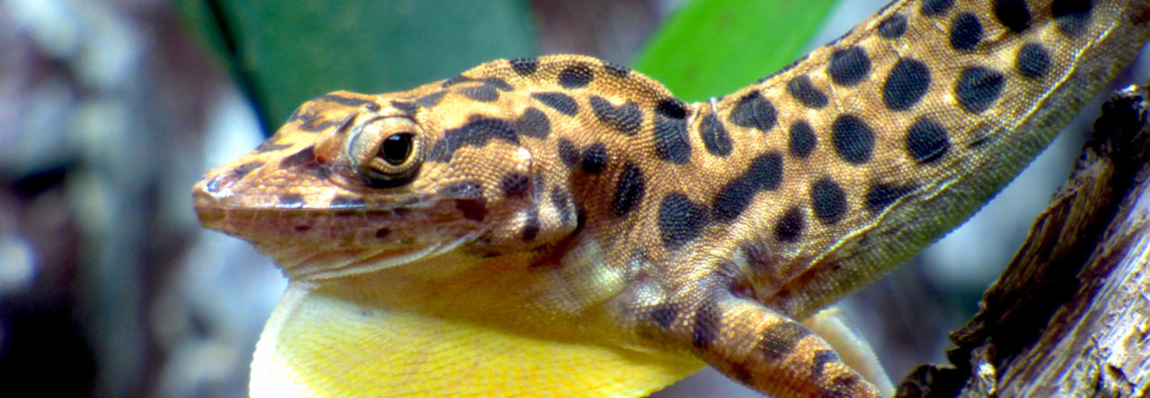
Photo from http://www.exoticpetvet.com/breeds/Green%20Anole.htm
Cornerstone recently reported abstracts from an undergraduate research symposium at the University of Minnesota Mankato. Included in the event were four projects from the laboratory of Rachel Cohen.
Seasonal Effects on Kisspeptin Concentration in the Green Anole Lizard, Anolis carolinensis
Nicholas Booker, Minnesota State University Mankato
Hyejoo Kang, Minnesota State University Mankato
Gonadal steroid hormones are responsible for reproductive behaviors; disruption in production of these hormones is also linked to fertility issues. The hypothalamic-pituitary- gonadal (HPG) axis controls the production of sex steroid hormones, testosterone and estradiol. A peptide, kisspeptin, stimulates this axis by acting on neurons in the hypothalamus. The green anole lizard, Anolis carolinensis, is a seasonally breeding animal that shows drastic changes in behavior and physiology between the breeding and non- breeding seasons. One such change is a large increase in testosterone levels in the breeding season compared to the non-breeding season. These fluctuations in testosterone concentration in green anoles allows for a great opportunity to study the HPG axis. In the current study, we used brain tissue from breeding and non-breeding season green anoles to perform western blot analysis on kisspeptin concentration. Due to the increase in testosterone in the breeding season, we hypothesized that an increase in kisspeptin concentrations will be observed in breeding season compared to the non-breeding season lizards. These results would suggest that kisspeptin does indeed play a role in stimulating the HPG axis and that kisspeptin could potentially be used as a treatment for infertility.
The Effect of Steroid Hormones on Neuronal Size and Number in Two Brain Regions Important for Reproduction
Jaeyoung Son, Minnesota State University Mankato
Steroid hormones, such as testosterone (T) and its metabolites (estradiol, E2, and dihydrotestosterone, DHT), are critical for the production of reproductive behavior. These hormones play a role in neural plasticity, such as changes in neuronal size change and brain region volume. Our study is examining the role of steroid hormones in maintaining the morphology of brain areas involved in reproduction, such as the ventromedial hypothalamus (VMH) and preoptic area (POA). We are using the green anole lizard (Anolis carolinensis) as a model because they are seasonally dimorphic, with more reproductive behaviors and higher steroid hormones in the breeding compared to non-breeding season. We treated our animals with different steroid hormones: T, DHT, E2, and blank capsules as a control. We collected the brains, sectioned the tissue and measured neuron size, number and density in the VMH and POA. We are expecting to find smaller and increased numbers of neurons in the animals treated with steroid hormones compared to the controls. This result would support the idea that steroid hormones are critical for the maintenance of brain areas important for reproduction.
Seasonal Variation in the Dorsolateral and Medial Cortex of Green Anole Lizards
Amber Day, Minnesota State University Mankato
Abdi Abdilahi, Minnesota State University Mankato
The hippocampus is a region of the brain involved in spatial learning and memory, and has been shown to add new neurons in adult animals. Steroid hormones, specifically testosterone
(T) and its metabolites (estradiol, E2, and dihydrotestosterone, DHT), have been shown to play a role in the addition of adult-born neurons to the brain. The green anole lizard, Anolis carolinensis, is a seasonally breeding animal that exhibits seasonally dimorphic behaviors, as well as seasonal anatomical differences in the brain. The pronounced differences between the breeding and non-breeding seasons make this lizard an excellent model for the study of how steroid hormone differences impact the brain. We examined the volume of and addition of new adult-born neurons to the dorsolateral and medial cortex in the lizard, which is analogous to the mammalian hippocampus. We sectioned brain tissue from breeding and non-breeding animals, performed a Nissl stain, and are measuring volume of the regions. We expect that the region will be larger in the breeding season due to the increase of territorial and courtship behaviors. We also treated animals with T, DHT, E2 or nothing as a control and performed an immunohistochemistry to examine how steroid hormones impact neurogenesis. We expect to see significantly more neurogenesis in the dorsolateral and medial cortex of T, DHT, E treated animals in comparison to the untreated group. Our experimental results may provide a greater understanding of the mechanisms that regulate the neural control of reproduction and territorial behaviors.
Amygdala Morphology and Neurogenesis in the Green Anole Lizard
Jadden Roddick, Minnesota State University Mankato
Nicholas Booker, Minnesota State University Mankato
Abodalrahman Algamdy, Minnesota State University Mankato
Steroid hormones and their derivatives play a major role in the reproductive system. One region in the brain that is involved in reproduction is the amygdala. We are examining the relationship between steroid hormones and neuron size, number and neurogenesis in the amygdala of the green anole lizard (Anolis carolinensis). Green anoles are exceptionally good models to examine the neural control of reproductive behaviors because they are seasonally breeding animals and exhibit unique behavioral and physiological differences in the breeding season compared to the non-breeding season. These behavioral differences are likely caused by seasonal changes in circulating steroid hormone levels. For our project, breeding green anole males were gonadectomized and a capsule containing testosterone, estradiol, dihydrotestosterone or left empty was inserted under the anole’s skin. The animals were injected with bromodeoxyuridine (BrdU; a new cell marker) for three days after the treatment. After one month, brains were collected, sectioned, and placed on slides. An immunohistochemistry for BrdU and Hu (neuronal marker) was conducted to examine the presence of new neurons in the amygdala. Alternate sections were Nissl stained and used to count cell number and measure soma size. We expect to see a decrease in neuron number, soma size, and neurogenesis in the animals treated with hormones compared to the animals treated with the blank capsule because we see this pattern in breeding season animals. This work will help provide more insight into the neural control of reproduction.
- Evolution in Real Time on Lizard Island - March 23, 2025
- Spider Snags Adult Anolis osa - March 22, 2025
- An Homage to the Green Anoles of New Orleans - March 21, 2025


Leave a Reply