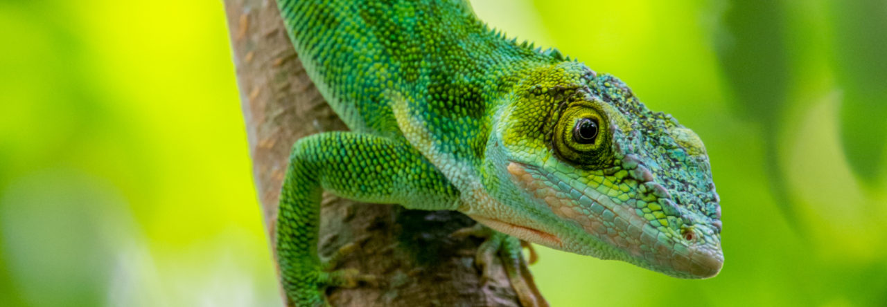
Over the last decade the term “model species” has taken on new meaning. Species that were once the building blocks for distinct disciplines have taken on new importance in comparative evolutionary studies that integrate perspectives across biological disciplines. Nowhere is this better illustrated than with Anolis lizards. For decades anoles were a workhorse of ecologists and evolutionary biologists, but have, more recently, been embraced by developmental biologists, genomicists, physiologists, and neurobiologists among others. This disciplinary expansion is perhaps most evident with the rapid increase of penis/hemipenis research that has been published using anoles within the year.
For many herpetologists, including those focused on anoles, the hemipenis is ripe with taxonomic characters, easily allowing for the identification of new species. Julia Klaczko and colleagues recently demonstrated that features of the hemipenis are some of the most rapidly evolving characters among anoles, a group already well known for its rapid anatomical evolution. Independent from these taxon-specific interests, developmental biologists became interested in the anole hemipenis because of its unique anatomy compared to other amniotes. Marissa Gredler and members of the Cohn Lab used anoles as one of their reptilian models of external genital development in what is arguably the broadest embryological survey of reptilian phallus development to date. In parallel, Patrick Tschopp and colleagues probed the cellular and molecular regulation of early phallus development among anoles, snakes, chickens and mice, demonstrating that the hemiphalluses (hemipenes and hemiclitores) and hindlimbs of squamates utilize similar molecular networks at the earliest embryonic stages of morphogenesis. Now, just within the last month, two more papers have used anoles in studies of phallus evolution and development, one using cutting-edge molecular techniques to better understand the relationship between limbs and external genitalia and the other addressing the fundamental question of external genital homology using museum specimens that are more than 100 years old.
Before getting into the findings of this new research, lets lay out some of the dirty details of penis evolution. First and foremost, the penises of amniotes are extremely diverse. Squamates have paired lateral phalluses while other clades have a single midline phallus. Each of the amniote lineages uses hydrostatic pressure to achieve an erection, yet accomplish this using different bodily fluids (lymph or blood). In mammals sperm is transferred to the female through a closed urethral tube, but other groups utilize an open channel. Most birds (97%) and the tuatara, have absent or highly reduced phalluses and reproduce with the famed “cloacal kiss.” These large differences in anatomy should not overshadow the spines, bulges, corkscrews, and dramatic differences in size that give species their distinctive features. But with such striking variation, we are forced to wonder how many times the penis evolved. Perhaps the amniote ancestor possessed an intromittent phallus capable to transferring sperm to the female that later diversified in each lineage independently. Or, perhaps the last common amniote ancestor used cloacal apposition to foster internal fertilization and unique phallus morphologies evolved independently at the origin of each lineage. Because adult anatomy provides few clues to phallus homology, Thom Sanger (me), Marissa Gredler, and Marty Cohn looked towards the embryo for help.

Table 1 from Sanger et al. 2015 summarizing phallus variation in amniotes
The tuatara, a species lacking an adult phallus, has presented a problem in attempts to reconstruct the last common ancestor of amniotes because it raises the distinct possibility that reproduction through cloacal apposition was the ancestral condition. However, in their previous survey of reptilian phallus development, the Cohn lab discovered that nearly all amniotes initiate genital development with paired swellings adjacent to the cloaca. To address whether the tuatara might have this signature of genital initiation, we utilized a unique museum collection of embryonic tuatara embryos collected over 100 years ago by Arthur Dendy (to this day still most well known for his taxonomy of sponges). At some point prior to 1907 Dendy sent Charles Sedwick Minot, “master histologist” and curator of the Harvard Embryological Collection, a series of four tuatara embryos that Minot histologically sectioned and stained. To the best of our knowledge, these embryos were never published on until now, but instead collected dust in storage since Minot’s death in 1917. But lo and behold, after piecing the embryo back together again through a computationally intensive version of humpy-dumpty, the same embryonic swellings could be found on the tuatara embryo as all other amniote species examined to date. It was actually somewhat shocking to see just how similar the tuatara reconstruction was to the genital swellings of the green anole. This observation suggests that the tuatara initiates genital development as an early embryo, but that it is lost before developing into a fully functional phallus. It also suggests that the phallus evolved only once at the origin of amniotes and that the variation observed in adult form arose through lineage specific modifications to those developmental programs. This research recently appeared in Biology Letters.

Reconstruction of a Sphenodon embryo (left) compared to an green anole embryo (right). (HL = hindlimb, AS = anterior swelling, GS = genital swelling). Modified from Sanger et al (2015).
Because of the mechanics of creating an enclosed amniotic egg, internal fertilization must be associated with the origin of amniotes. This allows us to hypothesize why the phallus evolved in the last common ancestor of amniotes, but addressing how the phallus evolved as an anatomical novelty requires a different approach.
For over a decade, developmental biologists have known of molecular similarities between the embryonic limbs and genitalia. Carlos Infante, Doug Menke and colleagues, therefore, hypothesized that the developing limbs and genitalia would also share a battery of enhancers– the non-coding on/off switches that control the proper time and place of gene expression. Furthermore, they hypothesized that if limbs and genitals have common regulatory landscapes early in development for most species, then these small bits of DNA may diverge in limbless species such as snakes following limb loss. To test these hypotheses, they set out to find, capture, and catalog the repertoire of enhancers present in early limb and phallus development in both limbed and non-limbed species. Using powerful computational methods, they described thousands of enhancers that are active in the early stages of limb and phallus development in limbed and non-limbed species, but found a more complex evolutionary scenario than they expected.
Within mice, there is substantial overlap in the enhancers powering the development of limbs and the penis. Genomic comparisons among vertebrates, including anoles, show that enhancers cataloged by their known role in (mammalian) limb development or those active in both limbs and genitalia show higher sequence conservation than enhancers involved only in external genitalia development. This suggests that there may be some selection pressure to maintain these elements, even after limb loss. When the authors looked to see what enhancers were actually functionally active in the developing limbs and genitalia of anoles, they found that limbs have a more conserved regulatory landscape than the external genitalia, likely as a result of their distinct mature anatomies. The authors move beyond broad, genome-wide comparisons to dissect the precise function and evolution of a pair of enhancers (HLA and HLB) for the gene Tbx4, a gene expressed in the early limbs and phallus. The activity of both of these enhancers is conserved between Anolis and mouse; HLA being expressed in limbs while HLB is expressed in both the limbs and external genitalia. But when the snake sequence of HLB is picked up and put into a mouse embryo, it was found to no longer drive limb expression, only genital expression. A portion of this enhancer’s function has evolved. Because of the widespread conservation of enhancers in the snake genome the authors conclude by speculating that this persistence may be due to additional enhancer functions in other urogenital and reproductive organs. This report was recently published in Developmental Cell.
In a parallel commentary the authors suggest that “future studies would benefit from a strategy that includes Anolis (instead of mouse) as a reference species” for functional studies. Now, wouldn’t that be an exciting day that we can all hope to witness in our lifetimes.
- Short Faces, Two Faces, No Faces: Lizards Heads Are Susceptible to Embryonic Thermal Stress - December 15, 2021
- The Super Sticky Super Power of Lizards: a New Outreach Activity for Grade-Schoolers - April 9, 2018
- Updates on the Development of Anolis as a “Model Clade” of Integrative Analyses of Anatomical Evolution - September 4, 2017


Leave a Reply